Alpha-MSH And The Melanocortin System: Your Body's Built-In Anti-Inflammatory Switch
By Jacob Gordon, INHC, FMT-CThis article contains affiliate links. As an Amazon Associate, MyBioHack earns from qualifying purchases at no extra cost to you. We only link products we research and stand behind.
Your body makes a hormone that calms inflammation, induces immune tolerance, protects your DNA from the sun, and guards the hair follicle and the eye from immune attack, all at once.
In this post, we will discuss where the melanocortins come from, what each of the five receptors does, how alpha-MSH shuts down inflammation, why the redhead receptor is a DNA repair gene, what the CIRS literature actually shows about low alpha-MSH, and which melanocortin drugs are real medicine versus a gray-market gamble.
---
- The Body's Built-In Anti-Inflammatory
- POMC: One Precursor, Many Hormones
- The Five Melanocortin Receptors
- How Alpha-MSH Shuts Down Inflammation
- KPV And The Melanocortin Tripeptides
- Alpha-MSH Builds Immune Tolerance
- MC1R, Pigment, And UV Protection
- MC3R, MC4R, And Energy Balance
- The Melanocortin System In The Gut, Joints, And Eye
- Agouti And AgRP: The Endogenous Antagonists
- Fever, Thermoregulation, And The Stress Axis
- The Thread Through The Whole Series
- The Junction Dysfunction And CIRS Connection
- Melanocortin Drugs And The Melanotan Problem
- How To Support The Melanocortin System
- Testing
- Mechanisms Of Action
- Genetics
- More Research
The Body's Built-In Anti-Inflammatory
Most anti-inflammatory drugs come from a pharmacy.
Your body already makes one of the most elegant anti-inflammatory molecules known.
Alpha-melanocyte-stimulating hormone (alpha-MSH) is a 13-amino-acid peptide that suppresses inflammation, induces immune tolerance, protects against oxidative and UV damage, and does it potently at very low concentrations. R
It belongs to a family of signals called the melanocortins, and the whole system is one of the body's master brakes on inflammation.
I use the word brake deliberately.
Alpha-MSH does not shut the immune system off the way a steroid does, it lowers the gain on the response and then rebuilds the regulatory cells that keep the gain low afterward. R
This is not a fringe molecule.
The melanocortin receptors are an active pharmaceutical target precisely because alpha-MSH does naturally what anti-inflammatory drugs try to do artificially. R
It has already appeared in every post in this series, as the guardian of hair follicle immune privilege, the anti-inflammatory melanocortin in sebocytes, and the protector of pigment cells in vitiligo.
This post is about the system itself.
POMC: One Precursor, Many Hormones
The melanocortins all come from one parent protein.
Proopiomelanocortin (POMC) is a large precursor protein that gets cut into several bioactive peptides: adrenocorticotropic hormone (ACTH), the melanocyte-stimulating hormones (alpha, beta, and gamma-MSH), and beta-endorphin. R
Which peptides you get depends entirely on which scissors the tissue owns.
The cutting is done by the prohormone convertases (PC1/3 and PC2), and their tissue-specific expression is what decides whether POMC becomes ACTH or gets processed further into alpha-MSH and beta-endorphin. R
In the anterior pituitary, PC1/3 dominates and the main product is ACTH, which is why the pituitary is a cortisol-driving organ rather than a pigment-driving one.
In the hypothalamus and in peripheral tissues, PC2 continues the job and clips ACTH down into alpha-MSH, and mice lacking PC2 or its chaperone 7B2 fail to generate the MSH peptides at all. R
Alpha-MSH is then finished by carboxypeptidase E and peptidyl-alpha-amidating monooxygenase, and that acetylation and amidation is what protects it from being chewed up instantly by peptidases. R
The final detail that matters clinically is that POMC is not a pituitary monopoly.
Skin, gut epithelium, immune cells, and the eye all transcribe POMC and make their own melanocortins locally, which means the anti-inflammatory signal is generated in the tissue that needs it rather than shipped in from the brain. R
That local production is the whole reason a skin condition or a colitis flare can have a melanocortin problem without any pituitary disease at all.
The Five Melanocortin Receptors
The melanocortins act on five G-protein-coupled receptors, MC1R through MC5R, and all five signal primarily by raising cyclic AMP. R
The roles break down roughly as follows (not an exclusive list):
- MC1R controls pigmentation and carries most of the anti-inflammatory signaling, expressed on melanocytes, keratinocytes, monocytes, macrophages, dendritic cells, mast cells, and endothelium
- MC2R is the ACTH receptor on the adrenal cortex, and it is the odd one out because it responds only to ACTH and not to any MSH peptide
- MC3R sits mostly in the hypothalamus and regulates the nutritional gate on growth and puberty
- MC4R sits in the hypothalamus and brainstem and is the dominant brake on appetite and the driver of energy expenditure
- MC5R regulates exocrine glands, including the sebaceous gland
MC1R is the star for skin and immunity because it has the highest affinity for alpha-MSH of the five and is expressed on exactly the cells that need calming. R
MC2R deserves a note because its oddity is instructive.
It cannot even reach the cell surface without an accessory protein called melanocortin receptor accessory protein (MRAP), and loss-of-function mutations in either MC2R or MRAP cause familial glucocorticoid deficiency, a condition where ACTH is sky high and cortisol is absent. R
MRAP2, its relative, does the same chaperone job for MC4R and is itself an obesity gene. R
MC5R was mapped to human sebaceous glands and turns out to be a marker of sebocyte differentiation, detected only in the lipid-laden differentiating cells and not the basal ones. R
That is the direct link between the melanocortin system and acne biology.
How Alpha-MSH Shuts Down Inflammation
The mechanism is clean and it converges on one node.
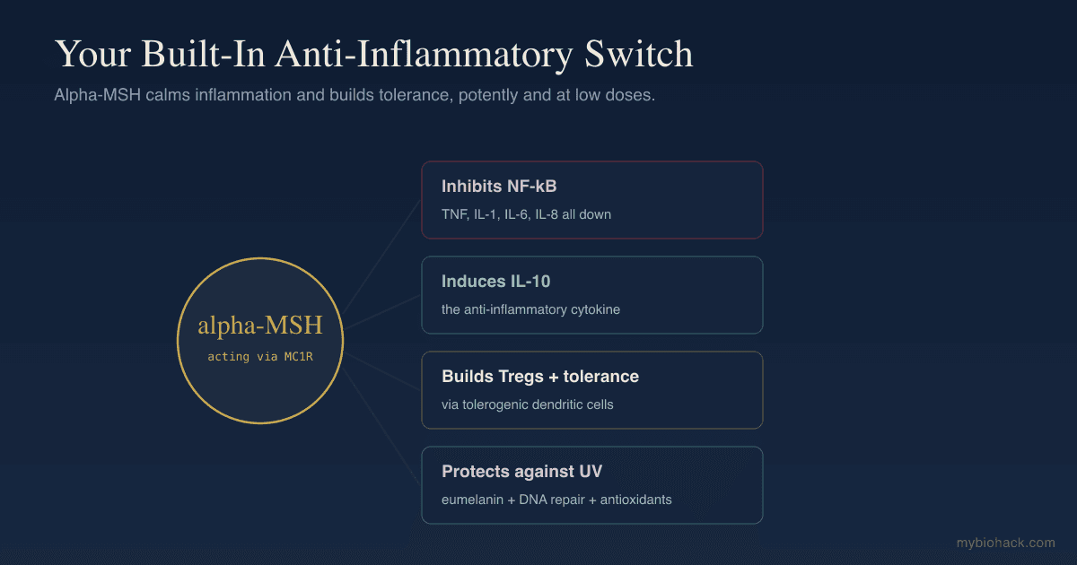
Alpha-MSH suppresses the production of pro-inflammatory cytokines including TNF-alpha, IFN-gamma, IL-1, IL-6, and IL-8, while inducing the anti-inflammatory cytokine IL-10. R
It does this largely by inhibiting nuclear factor kappa B (NF-kB), the master switch that turns on most inflammatory genes.
In human keratinocytes, alpha-MSH and ACTH both blocked TNF-alpha-stimulated nuclear translocation of NF-kB p65 in a dose-dependent way, which is the upstream event that all the downstream cytokine suppression follows from. R
The same suppression holds in dermal fibroblasts, where MSH peptides inhibited TNF-alpha signaling directly. R
Alpha-MSH also works upstream of NF-kB on the pattern recognition side.
Pretreating human keratinocytes with alpha-MSH downregulated TLR2 and IL-8 responses to staphylococcal lipoteichoic acid, and it did so by inducing IRAK-M, a negative regulator of TLR signaling, an effect that was abolished by the MC1R antagonist agouti signaling protein. R
That is an important detail, because it means alpha-MSH is not only quieting the cytokine output, it is raising the threshold at which the cell decides to respond to a microbial signal at all.
On the oxidative side, alpha-MSH reduced the damage done to keratinocytes and melanoma cells by pro-inflammatory cytokines and peroxide-generated oxidative stress. R
It also suppresses inducible nitric oxide synthase, reduces the adhesion molecules that recruit immune cells to a tissue, and dampens immune cell migration. R
Mast cells are in the circuit too.
Murine mast cells express MC1R, and alpha-MSH inhibited IgE-triggered histamine release by roughly 60 percent while downregulating IL-1beta, TNF-alpha, and lymphotactin transcripts. R
Anyone working on mast cell activation should sit with that number for a moment.
There is a genuine complication here that most write-ups omit.
Intradermal alpha-MSH also provokes scratching in mice through keratinocyte-derived histamine acting on H1 receptors, so the same peptide that calms mast cells systemically can be locally pruritogenic in the skin. R
That is a real contradiction, it is dose and route dependent, and it belongs in any honest account of the neuroscience of itch.
KPV And The Melanocortin Tripeptides
One of the most interesting discoveries in this field is that you do not need the whole alpha-MSH molecule.
The anti-inflammatory activity is concentrated in the last three amino acids, a tripeptide called KPV (lysine-proline-valine). R
KPV is fascinating because it works through a different door.
Rather than going through the melanocortin receptors, KPV is taken into immune and intestinal epithelial cells by the peptide transporter PepT1, and once inside, nanomolar concentrations inhibit NF-kB and MAP kinase inflammatory signaling. R
That receptor-independent route is why KPV is orally active and gut-directed rather than systemic and pigmentary.
Related fragments behave the same way, and the MSH 11-13 sequence signals in human keratinocytes alongside full-length alpha-MSH and ACTH. R
Oral KPV reduces the severity of chemically induced colitis in animal models and prevents colitis-associated cancer through PepT1. R
Delivering KPV in hyaluronic-acid-functionalized nanoparticles targeted it further to inflamed colon tissue and alleviated ulcerative colitis in mice at very low doses. R
All of that is rodent work.
Human clinical evidence for KPV is still essentially absent, and I am not going to pretend a mouse colitis model is a trial.
It is the anti-inflammatory essence of alpha-MSH shrunk to three amino acids, and it is a promising tool with a thin human file, in the same category as GHK-Cu on the repair side.
Alpha-MSH Builds Immune Tolerance
Beyond suppressing inflammation, alpha-MSH actively builds tolerance.
Acting through MC1R, alpha-MSH induces tolerogenic dendritic cells, the kind of antigen-presenting cell that calms the immune system rather than activating it. R
Those tolerogenic dendritic cells then expand regulatory T cells (Tregs), the CD4+CD25+Foxp3+ cells that hold autoimmune and allergic responses in check. R
These are functional cells, not just markers on a flow plot.
Alpha-MSH-induced Tregs inhibited contact allergy and calmed ongoing psoriasis-like skin inflammation in mice, and the same alpha-MSH-stimulated dendritic cells generated Tregs from human blood that suppressed Th17 cells taken from people with psoriasis. R
Part of the mechanism upstream of that is annexin A1.
Alpha-MSH inhibited TNF-alpha-induced maturation of human dendritic cells by upregulating ANXA1, which is the same pro-resolving protein that glucocorticoids work through. R
Alpha-MSH suppressed antigen-induced lymphocyte proliferation in human volunteers, and it did so independently of MC1R genotype, which points at more than one receptor carrying the immune signal. R
Alpha-MSH is also one of the mediators of UV-induced immunosuppression, which is the same tolerance machinery viewed from a less flattering angle. R
This tolerance-building role is exactly why alpha-MSH is one of the immune privilege guardians that protect the hair follicle and the eye from autoimmune attack.
So the melanocortin system does two things at once.
It turns down active inflammation, and it builds the regulatory cells that prevent the next attack.
MC1R, Pigment, And UV Protection
The melanocortin system is also your primary sunlight defense.
When UV hits the skin it drives POMC and alpha-MSH production, which acts on MC1R to switch melanocytes from making reddish pheomelanin to making dark, protective eumelanin. R
That switch is the entire basis of tanning, and blocking MC1R signaling pushes production back toward pheomelanin, which is a poorer shield and a worse redox actor. R
But MC1R does far more than make pigment.
MC1R genotype determines the damage UV does to melanocytes, with functional receptors enhancing DNA repair and antioxidant defenses and loss-of-function receptors leaving the cell exposed. R
The pigment arm and the repair arm are separable.
Downstream of MC1R, cAMP signaling splits into distinct pathways, one driving melanogenesis and another driving nucleotide excision repair, which is why you can in principle get the repair benefit without the tan. R
Pharmacologic cAMP induction applied topically both melanized skin and improved recovery from UV injury in MC1R-intact animals. R
Even melanoma-associated MC1R variants retain some redox signaling capacity that protects against oxidative DNA damage, which tells you the receptor is doing several jobs at once rather than one. R
The epidemiology follows the biology.
A meta-analysis of MC1R variants found consistently increased cutaneous melanoma risk carried by the loss-of-function alleles, with a measurable population attributable burden. R
The risk is not confined to obviously fair people, since a pooled analysis found MC1R variants raised melanoma risk in darker-pigmented Caucasians as well. R
MC1R also reaches into pain.
MC1R loss-of-function variants alter pain sensitivity and mu-opioid analgesic response in both mice and humans, which is the origin of the clinical folklore about redheads and anesthesia. R
A more recent review of that literature found the effect real but smaller and messier than the folklore suggests, with inconsistent findings across anesthetic classes. R
The practical takeaway is that MC1R genotype is a DNA repair and analgesia variable, not only a hair color one, and that is covered further in the sun damage pathways post.
MC3R, MC4R, And Energy Balance
The central melanocortin system is the most powerful appetite circuit humans have.
POMC neurons in the arcuate nucleus release alpha-MSH onto MC4R in the hypothalamus and brainstem, and that signal is what tells the body to stop eating and raise energy expenditure. R
Leptin sits directly upstream of it, which is why the pathway is usually called the leptin-melanocortin axis and why leptin resistance shows up as a melanocortin problem. R
MC4R deficiency is the most common single-gene cause of human obesity. R
Complete POMC deficiency in humans is rarer and more dramatic.
The original report described children with severe early-onset obesity, adrenal insufficiency from absent ACTH, and red hair pigmentation, which is the cleanest natural demonstration that one gene serves appetite, cortisol, and pigment at the same time. R
Later case series confirmed the same triad and showed how hard it is to treat by replacement alone. R
MC3R turned out to have a different job than MC4R.
Rare MC3R variants that reduce signaling delay puberty and reduce childhood growth, which positioned MC3R as the receptor that gates growth and reproductive timing on nutritional state. R
A large puberty-timing genetic study across the full allele frequency spectrum reinforced the melanocortin pathway's role in that timing. R
The Melanocortin System In The Gut, Joints, And Eye
The melanocortin system is not a skin and brain system with side gigs.
In the gut, MC1R is required for control of experimental colitis, and blocking the alpha-MSH to MC1R pathway worsens disease. R
Alpha-MSH also protected Caco-2 intestinal epithelial monolayers against cytokine-induced barrier damage, meaning it defends the tight junction itself and not only the immune cells sitting behind it. R
That is directly relevant to anyone working the gut-skin axis or chasing intestinal permeability.
In the joint, there is a full functional POMC system.
Synovial and bone cells make beta-endorphin, ACTH, and alpha-MSH, express MC1R, MC2R, MC3R, and MC4R, and melanocortin peptides show anti-inflammatory effects in animal models of arthritis. R
MC1R activation in synovial fibroblasts drove them into a senescent, non-inflammatory state that helped resolve arthritis rather than merely suppress it. R
Biased MC1R agonists have been designed specifically to capture that pro-resolving effect while avoiding the melanogenic one. R
In the eye, the ocular melanocortin system maintains the immune privilege of the anterior chamber and the retina, which is the same tolerance mechanism the hair follicle uses. R
Agouti And AgRP: The Endogenous Antagonists
A brake needs an accelerator, and the melanocortin system has two.
Agouti signaling protein (ASIP) is the peripheral antagonist that blocks MC1R on melanocytes, and blocking MC1R is what shifts pigment production toward pheomelanin. R
ASIP variants are established contributors to human pigmentation phenotypes including freckling, alongside MC1R itself. R
Agouti-related peptide (AgRP) is the central counterpart, released by a separate arcuate neuron population that opposes POMC neurons at MC3R and MC4R. R
AgRP is not a simple competitive blocker.
It behaves as an inverse agonist at MC4R, meaning it drives receptor signaling below baseline rather than just displacing alpha-MSH, which is why hunger signaling is so much stronger than a passive absence of satiety. R
The clinical lesson generalizes past appetite.
When a melanocortin signal looks low, the question is always whether you are producing less agonist or expressing more antagonist, and those are two different problems with two different fixes.
Fever, Thermoregulation, And The Stress Axis
Alpha-MSH is one of the body's endogenous antipyretics.
Given centrally, alpha-MSH suppressed LPS-induced fever in rats, and blocking MC4R not only prevented that effect but reversed it into an augmented fever. R
Alpha-MSH sits alongside glucocorticoids, arginine vasopressin, and other endogenous antipyretics as part of the built-in ceiling that keeps a fever from running away. R
Interestingly, the same study found that in afebrile animals alpha-MSH raised core temperature slightly, so the peptide is a thermoregulatory modulator rather than a simple cooler. R
The relationship to the HPA axis runs through the shared precursor.
POMC is the source of both ACTH and alpha-MSH, so pituitary stress output and melanocortin anti-inflammatory tone are drawn from the same pool and processed by the same convertases. R
The skin runs its own local version of the axis, with CRH, POMC, ACTH, and cortisol produced in situ and regulated by UV, which is why psychological stress reaches skin inflammation so quickly. R
Genetic variation in that cutaneous axis shapes how a given person's skin responds to UVB in the first place. R
If you want the fuller version of that circuit, it is in the stress and skin immune function post and the CRH resistance post.
The Thread Through The Whole Series
Step back and alpha-MSH is the quiet protagonist of this entire series.
It is the immune privilege guardian whose loss helps trigger alopecia areata.
It is the anti-inflammatory melanocortin in the sebocyte that opposes acne inflammation.
It is the pigment-cell protector whose failure contributes to vitiligo.
It induces the Tregs that calm psoriasis and contact allergy, and it modulates mast cell activity relevant to eczema. R
In every one of these conditions the brain-skin axis is tilting toward neurogenic inflammation, and the melanocortin system is the counterweight that should be pushing back.
When inflammation is chronic, that counterweight gets depleted or overwhelmed.
Restoring it, rather than only suppressing the inflammation, is the underused half of the strategy.
The Junction Dysfunction And CIRS Connection
This is also where the melanocortin system meets Jacob's Junction Dysfunction framework.
Low alpha-MSH is the marker most associated with the mold and biotoxin illness picture labeled CIRS, so it is worth being precise about what that literature actually shows.
The primary source is a time-series study of 21 people from water-damaged buildings, in which serum alpha-MSH was abnormally low and leptin abnormally high at baseline, both improved with cholestyramine, and symptoms returned within one to seven days on re-exposure. R
That study reported its own limitations plainly: participants were not randomly selected, there was no double-blinding, and no specific exposure agent was demonstrated. R
A follow-up clinical trial from the same group repeated the pattern in a larger group with the same design constraints. R
So here is my honest read.
The observation that alpha-MSH runs low in people with chronic multi-system inflammatory illness is credible and mechanistically coherent, because everything above tells you a chronically inflamed tissue is one that has already spent its melanocortin brake.
The inference that low alpha-MSH is the lesion, and therefore the thing to replace, is not supported by that evidence base, and it is a bigger claim than a small unblinded time series can carry.
Jacob's framing differs from the conventional CIRS model on exactly that point.
Jacob's hypothesis is that low MSH is a downstream marker of the chronic inflammation, glycocalyx damage, and stuck wound healing cycles that define Junction Dysfunction, which means the work is removing the drivers depleting the melanocortin system rather than dosing the hormone.
The upstream drivers he points at are glycocalyx degradation and the endotoxin load of what he calls Micro-Sepsis, described in the Junction Dysfunction and Micro-Sepsis chapter.
This is his hypothesis, not settled science, and I present it as such.
It is also why the overlap with long COVID and systemic long-term inflammation is worth taking seriously even though nobody has measured alpha-MSH properly in those populations.
Melanocortin Drugs And The Melanotan Problem
The melanocortin system has produced three approved drugs and one large unregulated market.
Afamelanotide is an alpha-MSH analog approved for erythropoietic protoporphyria, a genetic porphyria in which sunlight causes severe burning pain within minutes. R
A German observational cohort found afamelanotide improved sun tolerance and quality of life with an acceptable safety profile over long-term use. R
It has also been tested in vitiligo, where afamelanotide added to narrowband UVB produced faster and more complete repigmentation than phototherapy alone, at the cost of generalized darkening. R
Setmelanotide is an MC4R agonist approved for obesity caused by POMC or leptin receptor deficiency, where it produced substantial weight loss in an open-label trial in patients who had failed everything else. R
A later phase 3 trial extended it to children as young as two with MC4R pathway obesity. R
Note what setmelanotide is not.
It is a drug for rare monogenic disease, not a general obesity drug, and treating it as the latter is the single most common misreading of the melanocortin literature.
Bremelanotide is a non-selective melanocortin agonist approved for hypoactive sexual desire disorder in premenopausal women, covered in more detail in the PT-141 post.
The evidence here is weak, and I will say so.
An independent analysis of the RECONNECT phase 3 trials found that the validated efficacy outcomes produced effects ranging from nil to small, that eight of eleven prespecified outcomes had gone unpublished, and that the outcomes showing benefit were largely post hoc. R
The manufacturer-affiliated subgroup analyses read more favorably, which is exactly the tension you would expect. R
Then there is the gray market.
Melanotan I and melanotan II are sold online as tanning peptides, and a dermatology review documented cutaneous complications including darkening and change of existing moles, eruptive dysplastic nevi, and four case reports of melanoma emerging from existing moles during or shortly after use. R
Additional case reports include atypical melanocytic nevi after a single dose, priapism requiring intervention, renal infarction, and a possible link between melanotan II nasal spray and oral mucosal melanoma. R R R R
Dermatology bodies have flagged the unregulated use as a public health issue, and multiple national health agencies have issued warnings. R
None of that proves causation, and the reviews say so.
But you are looking at unregulated compounding, unknown purity, self-injection, and a mechanism that by design stimulates melanocyte proliferation in people who are usually taking it because their own MC1R signaling is poor.
I do not use these and I do not recommend them.
How To Support The Melanocortin System
The practical playbook is to stimulate the system safely and stop depleting it.
1. Use sunlight intelligently
UV is the natural stimulus for cutaneous POMC and alpha-MSH, so sensible, non-burning exposure supports the system, covered in the how to use the sun post.
Burning does the opposite, because it converts a hormetic signal into the exact oxidative and inflammatory load that depletes the brake.
The full alpha-MSH raising protocol, including the practical list I am deliberately not duplicating here, is in the dedicated alpha-MSH post.
2. Reduce the inflammation that depletes it
Chronic NF-kB-driven inflammation works against the melanocortin system, so the endotoxin, mast cell, and gut barrier strategies throughout this series protect it.
The NRF2 side of the redox response and the melanocortin side are complementary, not interchangeable.
3. Support cAMP signaling downstream of MC1R
Because all five melanocortin receptors signal through cyclic AMP, compounds that support that second messenger, like forskolin, are sometimes used to support melanocortin-type signaling. R
This is an indirect and non-specific approach, and I would not oversell it.
4. Cover the repair arm if your MC1R is weak
If you carry loss-of-function MC1R variants, you are down a DNA repair pathway, not only a tanning pathway.
Oral nicotinamide at 500 mg twice daily reduced new non-melanoma skin cancers by 23 percent over 12 months in a phase 3 randomized trial in high-risk patients, which is a repair-side intervention rather than a melanocortin one. R
Astaxanthin has reasonable human data for reducing UV-induced skin damage and supporting barrier function, though the trials are small. R
5. Consider the melanocortin peptides carefully
KPV and related fragments are investigational tools for gut and skin inflammation with strong rodent data and almost no human trial data, and they belong in a conversation with a knowledgeable practitioner rather than in a supplement stack. R
The tanning peptides are a different category entirely, and section 14 explains why I stay away from them.
6. Address the stress axis
Since POMC sits at the heart of the stress response and the skin runs its own local copy of that axis, a dysregulated stress axis changes melanocortin output directly. R
Nervous system work is not adjunctive here, it is upstream.
Testing
You can measure parts of this system, with real caveats.
Blood And Urine Markers
Alpha-MSH can be measured in serum through LabCorp, and it is one of the markers used in chronic inflammatory illness workups.
The limitations are significant and rarely stated.
Melanocortin peptides are small, rapidly cleared, and structurally similar to each other, and the pharmacokinetic work on synthetic analogs was driven in part by how quickly native alpha-MSH is degraded. R
There is no consensus reference range, assays differ between labs, and cross-reactivity with related POMC fragments is a real concern, so a single low value is a data point and not a diagnosis.
The downstream cytokines alpha-MSH normally suppresses (TNF-alpha, IL-6, IL-1 beta, IL-8) give a better picture of the inflammatory load the system is fighting than the peptide itself does.
Functional Lab Panels
I use the Immune Zoomer (Vibrant Wellness) to assess the broader inflammatory and autoimmune picture that reflects how much melanocortin brake is being spent.
I use the Hormone Zoomer (Vibrant Wellness) to assess the POMC-related stress hormone axis, and the DUTCH Complete (Precision Analytical) when I want the cortisol rhythm rather than a single point.
For the toxin and mold drivers most often associated with low MSH, I use the Toxin Zoomer (Vibrant Wellness), or the Mycotoxins Profile (RealTime Labs) when mold specifically is the question.
Because the gut is where MC1R protects the barrier, I use the Gut Zoomer (Vibrant Wellness) or the GI-MAP (Diagnostic Solutions) to assess the endotoxin and permeability side.
Genetics
MC1R variant status is worth knowing if you burn easily, and it shows up in general skin pigmentation genotyping rather than in a standard panel. R
Mechanisms Of Action
Simple:
- Your body makes a hormone called alpha-MSH that turns down inflammation and teaches the immune system to tolerate rather than attack, and it also protects your skin from the sun.
- When inflammation is chronic, this natural brake gets worn down, which is part of why so many skin and gut conditions persist.
Advanced:
- NF-kB inhibition. Alpha-MSH, primarily via MC1R and cAMP, blocks TNF-alpha-stimulated nuclear translocation of NF-kB p65 dose-dependently in human keratinocytes, suppressing the downstream cytokine, chemokine, and adhesion molecule program. R
- TLR threshold raising via IRAK-M. Alpha-MSH pretreatment induces IRAK-M in human keratinocytes, downregulating TLR2 and IL-8 responses to bacterial lipoteichoic acid, an effect abolished by the MC1R antagonist agouti signaling protein. R
- Annexin A1 induction. Alpha-MSH upregulates ANXA1 to inhibit TNF-alpha-driven maturation of human dendritic cells, converging on the same pro-resolving protein that glucocorticoids use. R
- Tolerance induction. MC1R activation by alpha-MSH generates tolerogenic dendritic cells that expand functional Foxp3+ Tregs, suppressing contact allergy and psoriasiform inflammation and inhibiting Th17 cells from psoriasis patients. R
- Mast cell stabilization. MC1R on mast cells raises cAMP biphasically, inhibiting IgE-mediated histamine release by roughly 60 percent and downregulating IL-1beta, TNF-alpha, and lymphotactin transcripts. R
- PepT1-mediated tripeptide action. The C-terminal tripeptide KPV is transported into immune and epithelial cells by PepT1 and inhibits NF-kB and MAPK signaling at nanomolar concentrations, independent of melanocortin receptors, making it orally active for gut inflammation. R
- Epithelial barrier protection. Alpha-MSH preserves tight junction integrity in Caco-2 intestinal monolayers against cytokine-induced damage, and MC1R is required for control of experimental colitis in vivo. R R
- UV cytoprotection with separable arms. MC1R signaling drives eumelanin synthesis, nucleotide excision repair, and antioxidant defense through partly divergent cAMP branches, so repair and pigmentation are dissociable and loss-of-function variants impair both. R R
- Central energy balance. Arcuate POMC neurons release alpha-MSH onto MC4R to suppress feeding and raise energy expenditure, opposed by AgRP acting as an inverse agonist that drives MC4R signaling below basal. R R
- Endogenous antipyresis. Centrally administered alpha-MSH suppresses LPS-induced fever through MC4R, and MC4R blockade reverses the effect into fever augmentation. R
- Pro-resolving synovial senescence. MC1R activation in synovial fibroblasts induces a senescent non-inflammatory phenotype that facilitates resolution of arthritis rather than suppression alone. R
Genetics
Several genes set melanocortin tone.
MC1R
MC1R encodes the melanocortin 1 receptor, the highest-affinity alpha-MSH receptor, expressed on melanocytes, keratinocytes, and most melanocortin-responsive immune cells.
The loss-of-function red-hair-color variants R151C (rs1805007), R160W (rs1805008), and D294H (rs1805009) reduce cAMP signaling, shifting pigment toward pheomelanin and weakening both UV protection and anti-inflammatory tone. R
rs1805007 and rs1805008 are among the variants most consistently associated with cutaneous melanoma risk in meta-analysis. R
The same loss-of-function variants alter pain sensitivity and mu-opioid analgesic response, an effect that is real but smaller than the clinical folklore claims. R R
Carrying these variants means a weaker DNA repair response to UV, not only a poorer tan. R
POMC
POMC encodes the precursor for ACTH, the MSH peptides, and beta-endorphin.
Rare biallelic loss-of-function variants cause severe early-onset obesity, ACTH-deficient adrenal insufficiency, and red hair, the triad that first proved one gene serves appetite, cortisol, and pigment. R
Heterozygous carriers show a milder obesity phenotype, and replacement strategies aimed at the ACTH arm alone did not correct the weight problem. R
MC4R
MC4R encodes the hypothalamic receptor that alpha-MSH uses to suppress appetite and raise energy expenditure.
Heterozygous loss-of-function variants make MC4R deficiency the most common monogenic form of human obesity, presenting as hyperphagia with accelerated linear growth in childhood. R
MC4R also mediates the antipyretic effect of central alpha-MSH, so the same receptor gates appetite and fever. R
MC3R
MC3R encodes the second central melanocortin receptor and behaves as the nutritional gate on growth and puberty rather than a satiety receptor.
Rare signaling-reducing variants delay puberty and reduce childhood growth and adult height. R
MC2R And MRAP
MC2R encodes the adrenal ACTH receptor, the only melanocortin receptor that does not respond to MSH peptides.
Loss-of-function mutations in MC2R or in its obligate trafficking partner MRAP cause familial glucocorticoid deficiency, with very high ACTH and absent cortisol. R
MRAP2 performs the same chaperone role for MC4R and is itself implicated in obesity. R
MC5R
MC5R encodes the exocrine gland receptor and is detectable only in differentiating, lipid-laden human sebocytes, making it a marker of sebocyte differentiation and the direct link from melanocortins to sebum. R
ASIP
ASIP encodes agouti signaling protein, the endogenous MC1R antagonist that opposes alpha-MSH at the receptor and shifts melanogenesis toward pheomelanin. R
ASIP variants contribute to freckling and pigmentation phenotypes alongside MC1R. R
PCSK1
PCSK1 encodes prohormone convertase 1/3, the enzyme that makes the first cut in POMC.
Humans and mice with inactivating PC1/3 mutations are obese, which was long assumed to be a POMC processing failure. R
That assumption did not hold, because selectively deleting PC1/3 only in POMC-expressing cells does not produce obesity, which means the convertase phenotype runs through non-POMC peptides as well. R
More Research
A few additional threads are worth following.
Alpha-MSH and melanocortin analogs are protective across a wide range of animal models including uveitis, colitis, arthritis, and experimental autoimmune encephalomyelitis, which is why the melanocortin receptors are an active drug target well beyond dermatology. R
Biased agonism is the most interesting near-term development, because MC1R ligands can be designed to keep the pro-resolving immune effect while dropping the melanogenic one, which would sidestep the tanning problem entirely. R
Dersimelagon, an oral non-peptide MC1R agonist, showed disease-modifying effects in preclinical systemic sclerosis models, which extends the melanocortin story into fibrosis. R
MC1R's role in DNA repair and antioxidant defense means it is a cancer-relevant gene as much as a pigmentation gene, which reframes red-hair variants as a repair issue rather than a cosmetic one. R
The KPV route through PepT1 rather than melanocortin receptors opens a separate, orally available anti-inflammatory path that does not touch pigmentation at all, and it is the part of this field I would most like to see tested properly in humans. R
The unresolved question I keep coming back to is whether low alpha-MSH in chronic illness is ever primary, because nobody has run the study that would answer it: a properly blinded cohort with standardized assays, matched controls, and repeated measures across a treatment that fixes the upstream inflammation.
For the practical protocol see the alpha-MSH post, for the broader framework the brain-skin axis pillar, and for the glycocalyx side of the JD argument the improving the glycocalyx chapter.
For biomarker testing I use the Immune Zoomer and the Toxin Zoomer to assess the inflammatory load and toxin drivers that deplete the melanocortin system.
If you want help working with your melanocortin system instead of only suppressing inflammation, reach out for a consultation.
Jacob Gordon
INHC, FMT-C
Integrative Nutrition Health Coach
I cover mold illness, post-viral recovery, methylation, and complex chronic disease, drawing on ten years of clinical research, work inside a functional medicine clinic, and my own recovery from all of it. Every claim here is cited.
Book a ConsultationRelated Protocols & Supplements
Deep-dive chapters and recommended supplements for this topic
Quercetin
500mg 2x/day
SPM Active (Pro-resolving Mediators)
2 softgels/day
Curcumin (Liposomal)
500mg 2x/day