Mast Cells, Substance P, And Neurogenic Inflammation: The Nerve-To-Mast-Cell Loop Behind Flushing And MCAS
By Jacob Gordon, INHC, FMT-CThis article contains affiliate links. As an Amazon Associate, MyBioHack earns from qualifying purchases at no extra cost to you. We only link products we research and stand behind.
Most mast cell reactions are blamed on allergies, but your own nerves can fire mast cells directly with no allergen involved at all.
In this post, we will discuss how sensory nerves and mast cells form a self-amplifying loop, why substance P is the nerve's direct line to the mast cell, how this drives flushing, hives, and MCAS-style reactions, and what actually calms the loop.

The Mast Cell Is A Neuroimmune Cell
The mast cell is usually introduced as an allergy cell, the thing behind hay fever and anaphylaxis.
That description badly undersells it.
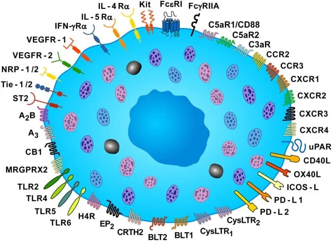
Mast cells are tissue-resident immune cells packed with granules of preformed histamine, tryptase, and tumor necrosis factor, positioned deliberately next to blood vessels and nerve endings. R
In humans, mast cells come in two main types based on their protease content.
The MCT type contains tryptase only and dominates mucosal surfaces like the gut and airway, while the MCTC type contains both tryptase and chymase and dominates connective tissues, especially the skin. R
Skin is MCTC territory, which matters because MCTC cells are the ones most responsive to neuropeptides and non-allergic triggers.
The placement is the point.
A cell loaded with inflammatory mediators, sitting on a blood vessel, wired to a sensory nerve, is not just an allergy cell.
It is a neuroimmune switch.
Two Ways To Trigger A Mast Cell

There are two fundamentally different ways to set a mast cell off.
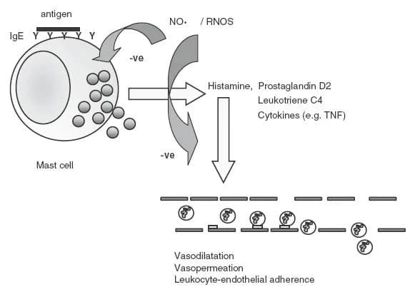
The first is the classic allergic route.
An allergen cross-links immunoglobulin E (IgE) antibodies bound to the mast cell, which triggers degranulation. This is true allergy, and it is what allergy testing looks for.
The second route does not involve IgE or antibodies at all.
Mast cells can be activated directly by cationic molecules, including your own neuropeptides, through a receptor called Mas-related G protein-coupled receptor X2 (MRGPRX2). R
This was a major discovery, because it means a mast cell can be fired by nerve signals, drugs, dyes, and toxins with no allergy present. R
This is the molecular basis of pseudo-allergic and neurogenic reactions.
It is also why so many people with clear-cut mast cell symptoms have completely negative allergy testing.
Their mast cells are not being triggered by allergens.
They are being triggered by their own nervous system.
Substance P: The Nerve's Direct Line
The single most important neuropeptide in this story is substance P (SP).
Substance P is released from sensory nerve endings in the skin, and human skin mast cells respond to it by releasing histamine in a dose-dependent way, a property that sets dermal mast cells apart from many other mast cell populations. R
Substance P hits mast cells through two receptors.
The classic one is the neurokinin-1 receptor (NK-1R), and the newer, often more powerful one is MRGPRX2. R
The downstream effects go well beyond histamine.
When substance P acts together with the alarm cytokine interleukin-33 (IL-33), it dramatically amplifies mast cell production of tumor necrosis factor and vascular endothelial growth factor (VEGF), and increases the vascular permeability of the skin. R
That last detail is the whole game.
Substance P, acting on the mast cell, makes blood vessels leak.
There is a subtle but important nuance in how this looks on skin.
A substance P antagonist blocks the spreading flare (the neurogenic vasodilation) but not the central wheal (the direct mast cell swelling), which tells you substance P drives both a nerve-mediated and a mast-cell-mediated component of the reaction. R
Neurogenic Inflammation: The Self-Amplifying Loop
Here is where it becomes a loop instead of a one-way signal.
When a sensory C-fiber fires, it releases substance P and calcitonin gene-related peptide (CGRP) backward into the skin through the axon reflex.
Those neuropeptides degranulate mast cells, and the mast cells release histamine, tryptase, and nerve growth factor (NGF). R
Then the mast cell mediators act back on the nerve.
Histamine, tryptase, and especially NGF sensitize the sensory nerve and even cause it to sprout new fibers, lowering the threshold for the next firing. R
This is neurogenic inflammation, and the mast cell and the nerve are so tightly coupled that researchers treat them as a single functional unit. R
The loop is self-reinforcing.
Nerve fires, mast cell degranulates, mediators sensitize and sprout the nerve, nerve fires more easily, and the cycle tightens.
This is the mechanistic reason chronic mast cell and skin conditions get worse over time and become reactive to smaller and smaller triggers. R
PAR-2 And Tryptase: The Mast Cell Talks Back
There is a second messenger in the mast-cell-to-nerve direction worth understanding on its own.
Mast cells release the enzyme tryptase, and tryptase activates a receptor on sensory nerves called proteinase-activated receptor 2 (PAR-2). R
PAR-2 activation on the nerve does two things.
It transmits itch directly, and it triggers the nerve to release even more substance P and CGRP, feeding the neurogenic loop. R
This pathway is especially important because it is histamine-independent.
In conditions like atopic dermatitis, tryptase is elevated up to fourfold and PAR-2 is upregulated on nerve fibers, which is a large part of why antihistamines so often fail to control the itch. R
So the mast cell and the nerve have two-way wiring.
The nerve talks to the mast cell through substance P.
The mast cell talks back through tryptase and NGF.
Stress, CRH, And The Mast Cell
The mast cell is also a direct target of stress hormones.
Corticotropin-releasing hormone (CRH), the master stress signal, activates dermal mast cells and triggers degranulation with increased vascular permeability. R
Acute psychological stress raises skin CRH, degranulates mast cells, and increases vascular leak, an effect that antihistamines can partly block. R
This is the molecular reason mast cell symptoms flare with stress even when nothing in the environment has changed.
For the full picture of how acute and chronic stress reshape skin immunity, see the companion post on stress and your skin.
The mast cell sits at the intersection of three inputs: allergens, nerves, and stress hormones.
In chronically ill people, the second and third inputs usually matter more than the first.
Why This Matters For MCAS And Flushing
This reframes a lot of confusing symptoms.
If you flush, itch, get hives, or react to heat, friction, emotion, exercise, or stress with no identifiable allergen, the nerve-MRGPRX2-mast cell loop is a leading explanation. R
This is the upstream layer that most Mast Cell Activation Syndrome and histamine intolerance protocols underweight.
They focus on stabilizing the mast cell and on degrading histamine, which is necessary, but they often skip the sensory nerve that keeps firing the mast cell in the first place.
If the nerve stays sensitized, the mast cells keep getting triggered no matter how many stabilizers you stack.
This is also why limbic and nervous-system work, which most people think of as unrelated to mast cells, can be one of the most effective interventions.
You are turning down the nerve that fires the cell.
The Junction Dysfunction Connection
Step back and look at what every trigger in this post produces.
Substance P makes vessels leak.
CRH makes vessels leak.
Mast cell degranulation makes vessels leak.
Increased vascular permeability is the shared endpoint, and in Jacob's Junction Dysfunction framework, leak at the microvascular level is Transient Capillary Leak Syndrome (TCLS). R
Jacob's framing is that the nerve-mast cell loop in the skin is one of the most visible examples of TCLS you can find, neurogenic signaling opening the capillaries and lymphatics.
There is a deeper layer here too.
In Jacob's model, mast cells in poorly perfused connective tissue degranulate partly because they are hypoxic, releasing histamine in an attempt to re-open and re-oxygenate the tissue, which is the subject of the JD chapter on mitochondria and mast cells in hypoxia.
So the mast cell is not malfunctioning at random.
It is responding to a stressed, hypoxic, nerve-driven environment exactly the way it evolved to.
For the full mechanism, see the TCLS chapter in the JD guide.
What Calms The Nerve-Mast Cell Loop
The key insight is that you have to address both ends of the loop, the mast cell and the nerve.
1. Stabilize the mast cell
Several natural flavonoids stabilize mast cell membranes and reduce mediator release.
The best studied are luteolin and quercetin, and the combination of palmitoylethanolamide and luteolin is particularly useful because it also calms the surrounding glia and nerves.
Vitamin C and stinging nettle are gentle additions, and prescription stabilizers like cromolyn and ketotifen exist for more severe cases.
2. Quiet the sensory nerve
This is the part most protocols miss.
Calming the nerve means reducing substance P and NGF signaling and desensitizing the sensory fibers.
Limbic retraining and nervous-system work directly reduce the central drive to fire these nerves, which is covered in the JD chapter on overcoming trauma's effect on the limbic system.
Beta-caryophyllene, a CB2-selective cannabinoid, reduces neurogenic inflammation without a psychoactive effect.
3. Degrade the histamine you do release
Supporting the enzymes that break down histamine reduces the symptom load.
This is the focus of the full histamine intolerance protocol, including DAO support and a lower-histamine diet.
4. Reduce the triggers that fire MRGPRX2 and the nerves
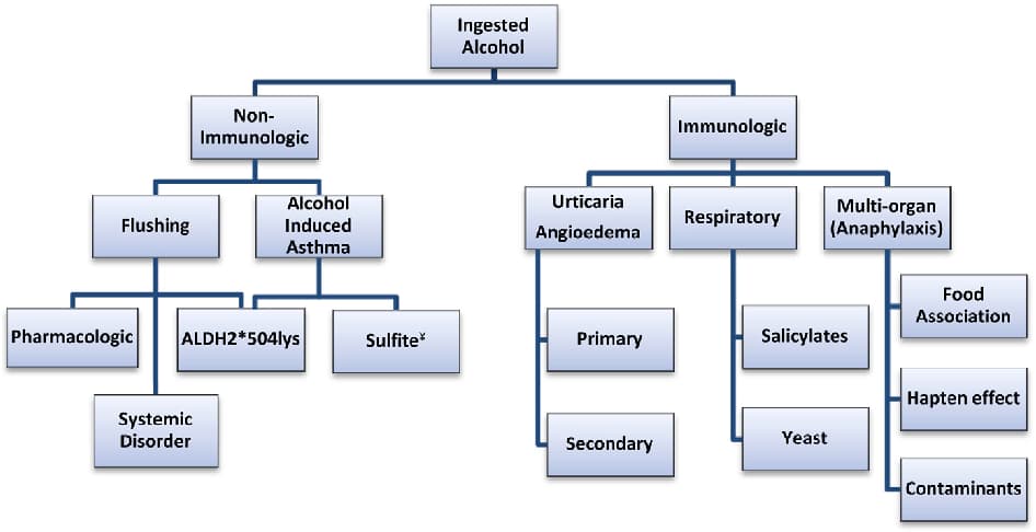
Common non-allergic triggers to reduce include alcohol, extreme heat and cold, friction, and rapid temperature swings, all of which can fire mast cells through the neurogenic route rather than through IgE.
Testing
You can objectively assess mast cell burden, though timing matters.
Blood And Urine Markers
Tryptase is the most stable mast cell marker and the standard for assessing total mast cell load.
Plasma histamine reflects active mast cell and basophil release but is short-lived, so it is best drawn during or near a reaction.
I use the Tryptase (Quest) and Plasma Histamine (Quest) tests to gauge mast cell activity.
Functional Lab Panels
For the broader picture including mast cell markers and autoimmune reactivity, I use the Immune Zoomer (Vibrant Wellness).
Because gut dysbiosis and histamine-producing bacteria feed this system, the Gut Zoomer (Vibrant Wellness) is often worth running alongside it.
Mechanisms Of Action
Simple:
- Your nerves release a chemical called substance P that makes nearby mast cells dump histamine, with no allergy involved.
- The mast cell then releases chemicals that make the nerve more sensitive, so the two keep firing each other in a loop that tightens over time.
Advanced:
- Dual-receptor activation. Substance P activates mast cells through both NK-1R (G-protein-coupled, NF-kB-driven cytokine transcription) and MRGPRX2 (IgE-independent, rapid degranulation), with skin MCTC mast cells being especially responsive. R
- Synergistic amplification. Substance P combined with IL-33 increases mast cell TNF and VEGF output by orders of magnitude and raises cutaneous vascular permeability, linking neuropeptide signaling directly to vascular leak. R
- The PAR-2 feedback arm. Mast-cell-derived tryptase cleaves and activates PAR-2 on sensory neurons, transmitting histamine-independent itch and stimulating further release of substance P and CGRP. R
- NGF-mediated sensitization. Mast cell NGF sensitizes and sprouts peptidergic nerve fibers, lowering activation thresholds and establishing the self-amplifying neurogenic loop. R
- CRH-driven degranulation. Skin CRH activates CRHR1 on dermal mast cells, producing selective, vascular-permeability-increasing degranulation, the effector arm of the cutaneous stress response. R
Genetics
Several genes set how reactive your mast cells and nerves are.
MRGPRX2
MRGPRX2 encodes the receptor for IgE-independent, neurogenic mast cell activation by substance P and basic secretagogues.
Higher expression or gain-of-function variation may explain why some people have far more pseudo-allergic reactivity. R
TPSAB1
TPSAB1 encodes alpha-tryptase, and extra copies of this gene cause hereditary alpha-tryptasemia, a common trait associated with elevated baseline tryptase, flushing, and heightened mast cell reactivity.
This is a copy-number trait rather than a single point mutation.
KIT
KIT encodes the receptor that controls mast cell survival and proliferation.
The somatic D816V mutation drives systemic mastocytosis, an expansion of mast cell numbers.
HNMT And AOC1
These genes encode the two enzymes that degrade histamine, histamine N-methyltransferase and diamine oxidase.
The HNMT Thr105Ile variant (rs11558538) reduces histamine-degrading activity.
Reduced activity means slower histamine clearance and a bigger symptom load, covered in the histamine intolerance post.
More Research
A few further threads are worth following.
Mast cell heterogeneity is more of a spectrum than two clean categories, and the connective-tissue MCTC mast cells of the skin appear especially tuned to neuropeptide and non-immune triggers. R
Substance P is broken down by peptidases in the skin, so the bioavailability of the signal depends on both how much is released and how fast it is degraded, which is why peptidase activity is an underappreciated variable in neurogenic inflammation. R
NK-1R antagonists, originally developed for other uses, are now being studied specifically for chronic itch and neurogenic inflammation, reflecting how central substance P is to this loop. R
For the receptor side of mast cell sensing, see the post on TRPV receptors, which are the heat, capsaicin, and chemical sensors that also feed this system.
For the broader framework, start with the brain-skin axis pillar.
For biomarker testing I use the Tryptase and Immune Zoomer to assess mast cell burden and reactivity.
If you have stubborn mast cell symptoms with negative allergy testing, reach out for a consultation.
Jacob Gordon
INHC, FMT-C
Board Certified Health Coach
I spent years battling unexplained chronic illness before discovering biohacking, epigenetics, and functional medicine. Now I share that research at MyBioHack to help others find their own answers.
Book a ConsultationRelated Protocols & Supplements
Deep-dive chapters and recommended supplements for this topic
Quercetin
500mg 2x/day
Vitamin D3 + K2
5000 IU + 200mcg/day
DAO Enzyme
1 cap before meals