Trigeminal Neuralgia: Causes, Mechanisms, Genetics, And How To Improve It
By Jacob Gordon, INHC, FMT-CThis article contains affiliate links. As an Amazon Associate, MyBioHack earns from qualifying purchases at no extra cost to you. We only link products we research and stand behind.
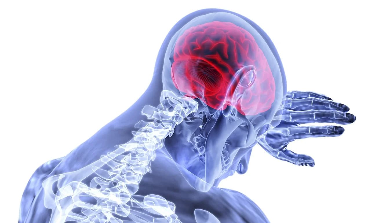
Trigeminal Neuralgia (TN) is a chronic neuropathic pain condition that produces sudden, severe, electric shock-like pain in the face, often triggered by something as innocuous as a breeze, a light touch, or chewing.
In this post, we will discuss the basics of trigeminal neuralgia, what causes it, how the trigeminal nerve works, overlapping conditions, how to improve it with supplements and lifestyle changes, what to avoid, relevant testing, mechanisms of action, genetics, and emerging research.

1. Basics Of Trigeminal Neuralgia
Trigeminal neuralgia is sometimes called tic douloureux (French for "painful twitch"), a name that dates back to the 18th century.
It is considered one of the most severe forms of pain a human being can experience.
The pain follows the distribution of the trigeminal nerve (cranial nerve V), the largest cranial nerve and the primary sensory nerve of the face. R
Attacks are typically unilateral (one side of the face) and last from a fraction of a second to about two minutes per episode. R
However, clusters of attacks can recur dozens to hundreds of times per day in severe cases.
TN is classified into three categories by the International Classification of Headache Disorders (ICHD-3):
- Classical TN is caused by neurovascular compression of the trigeminal nerve root, confirmed on MRI or during surgery
- Idiopathic TN presents with the same symptoms but without any identifiable cause on imaging
- Secondary TN is caused by an underlying disease such as multiple sclerosis (MS), tumor, or structural abnormality
The annual incidence of TN is estimated at 4 to 13 per 100,000 people, with more recent population studies suggesting around 5.5 per 100,000 person-years. R
Women are affected more than men, with a female-to-male ratio of approximately 3:2. R
Incidence increases sharply with age, rising from 0.1 per 100,000 in people under 20 to 23.1 per 100,000 in those over 80. R
The peak age group for prevalence is 51 to 59 years old. R
The right side of the face is affected more frequently than the left, at a ratio of approximately 1.5:1. R
Bilateral TN (both sides affected) is rare and typically raises suspicion for secondary causes like multiple sclerosis. R
Pain most commonly involves the V2 (maxillary) and V3 (mandibular) branches, either individually or combined.
Isolated V1 (ophthalmic) involvement is the least common, occurring in roughly 4% of cases. R
2. What Causes Trigeminal Neuralgia
The most common cause of classical TN is neurovascular compression (NVC), where a blood vessel (usually the superior cerebellar artery) presses against the trigeminal nerve root at its entry into the brainstem. R
This chronic pulsatile compression leads to focal demyelination (loss of the nerve's insulating sheath) at the root entry zone (REZ), the transition point between central and peripheral myelin. R
Demyelination exposes bare axons that then generate ectopic (abnormal) nerve impulses and allow ephaptic transmission (electrical crosstalk) between adjacent nerve fibers. R
This means a light touch signal traveling on an A-beta fiber can "jump" to an adjacent pain fiber (A-delta or C fiber), causing innocuous stimuli to trigger severe pain.
However, neurovascular compression alone does not explain all cases.
Up to 50% of classical TN patients show no clear morphological compression on imaging, and neurovascular contact is also found in many asymptomatic individuals. R
This suggests additional contributing factors:
- Autoimmune demyelination as seen in multiple sclerosis, where plaques in the brainstem directly damage the trigeminal pathways R
- Central sensitization, where repeated nociceptive input increases the excitability of second-order neurons in the trigeminal nucleus caudalis R
- Inflammatory cytokines such as IL-6 and TNF-alpha released at the compression site, which further sensitize the nerve R
- Satellite glial cell (SGC) activation in the trigeminal ganglion, driven by Toll-like receptors (particularly TLR3 and TLR5), producing inflammatory mediators R
- NLRP3 inflammasome activation in glial cells, which drives maturation of IL-1-beta and IL-18, amplifying neuroinflammation R
- Mitochondrial dysfunction in the injured trigeminal ganglion, spanning deficits in transcription, translation, and bioenergetic function R
- Chronic sinusitis and dental infections, which can inflame or compress the maxillary (V2) and mandibular (V3) branches of the trigeminal nerve through direct extension of infection or inflammatory cytokine release into the nerve territory R
- Post-infectious nerve injury from viral infections (particularly herpes simplex and varicella-zoster reactivation), which can damage trigeminal ganglion neurons and trigger persistent neuropathic pain R
- Tumors (particularly cerebellopontine angle tumors, meningiomas, and epidermoid cysts) compressing the nerve R
- Arteriovenous malformations and aneurysms near the trigeminal root
- Genetic channelopathies involving voltage-gated sodium and calcium channels (covered in the Genetics section)
A newer perspective suggests that some cases of TN may represent a primary demyelinating disorder rather than demyelination being secondary to vascular compression. R
This is worth keeping in mind, as it shifts the therapeutic focus from pure surgical decompression toward neuroprotective and remyelination strategies.
3. How The Trigeminal Nerve Works
The trigeminal nerve is the fifth cranial nerve (CN V) and the largest of the twelve cranial nerves.
It originates from the trigeminal nucleus complex in the brainstem and exits through the trigeminal ganglion (also called the Gasserian ganglion), a cluster of sensory nerve cell bodies located in Meckel's cave at the base of the skull.
From the ganglion, it divides into three branches:
- V1 (Ophthalmic) exits via the superior orbital fissure and supplies the forehead, upper eyelid, cornea, and bridge of the nose (purely sensory)
- V2 (Maxillary) exits via the foramen rotundum and supplies the cheek, upper lip, upper teeth, nasal cavity, and palate (purely sensory)
- V3 (Mandibular) exits via the foramen ovale and supplies the lower jaw, lower lip, chin, tongue, and ear region (mixed sensory and motor, controlling the muscles of mastication)
The sensory portion of the trigeminal nerve carries signals for touch, pain, temperature, and proprioception from the face to the brainstem.
The motor root travels exclusively with V3 and innervates the masseter, temporalis, medial pterygoid, and lateral pterygoid muscles used for chewing.
The root entry zone (REZ) is the critical region where the trigeminal nerve root transitions from peripheral myelin (produced by Schwann cells) to central myelin (produced by oligodendrocytes). R
This transition zone is structurally vulnerable because the central myelin segment is shorter and less resilient than peripheral myelin.
When a blood vessel compresses this zone, the oligodendrocyte-produced myelin degrades first, creating the conditions for ectopic firing and ephaptic crosstalk.
The trigeminal system also interacts extensively with the trigeminovascular system, which plays a central role in migraine and other headache disorders.
Trigeminal sensory neurons express TRPV1 receptors and release Calcitonin Gene-Related Peptide (CGRP), a neuropeptide that promotes vasodilation and neurogenic inflammation. R
CGRP potentiates TRPV1 activity, creating a feed-forward loop of sensitization in trigeminal neurons. R
Elevated CGRP in the upper spinal cord promotes sensitization of primary trigeminal nociceptive neurons through activation of protein kinase A (PKA) and phosphorylated extracellular signal-regulated kinase (P-ERK). R
4. Trigeminal Neuralgia And Overlapping Conditions
TN rarely exists in isolation, especially in patients with complex chronic illness.
Understanding the overlap with other conditions can reveal shared root mechanisms and open up additional treatment targets.
Multiple Sclerosis (MS)
The association between TN and MS is the most well-established comorbidity.
A systematic review and meta-analysis found the pooled prevalence of TN in MS patients to be approximately 3.4%, though individual studies report rates as high as 9.7%. R
The incidence of TN in MS patients is 15-fold higher than in the general neurological outpatient population. R
In MS-related TN, demyelinating plaques in the brainstem near the trigeminal root entry zone are the primary mechanism rather than vascular compression. R
TN commonly precedes the diagnosis of MS, making it a potential early warning sign. R
Neurovascular contact plays no significant role in TN secondary to MS. R
POTS And Dysautonomia
The presence of autonomic symptoms (tearing, nasal congestion, conjunctival injection) in TN correlates with worse surgical outcomes after microvascular decompression. R
Patients with Postural Orthostatic Tachycardia Syndrome (POTS) and other forms of dysautonomia share common pathways with trigeminal pain, particularly through small fiber neuropathy and autonomic nerve involvement.
The trigeminal autonomic reflex connects the trigeminal sensory system directly to parasympathetic autonomic output, meaning trigeminal activation can trigger measurable autonomic responses. R
Ehlers-Danlos Syndrome (EDS) And Connective Tissue Disorders
Patients with Ehlers-Danlos Syndrome (EDS), particularly the hypermobile type, are prone to craniofacial pain syndromes including trigeminal neuralgia.
Headache disorders in EDS patients are variably associated with cardiovascular dysautonomia, cervical spine and temporomandibular joint instability, meningeal fragility, poor sleep quality, and central sensitization. R
EDS, POTS, and mast cell activation frequently coexist as a recognized clinical triad. R
Mast Cell Activation Syndrome (MCAS)
Mast cells reside in brain parenchyma, meninges, and in close proximity to peripheral nerves, where they orchestrate neuroinflammatory processes. R
Degranulation of dural mast cells induces a prolonged state of excitation in meningeal nociceptors and downstream activation of the spinal trigeminal nucleus. R
Mast cell-glia crosstalk accelerates neuroinflammatory disease progression through release of cytokines, proteases, and reactive oxygen species. R
For more on this axis, see the post on mast cell-glia interaction.
SALI And Chronic Neuroinflammation
Systemic And Long-term Inflammation (SALI) provides a framework for understanding how TN fits into broader chronic illness.
Persistent glial activation, elevated cytokine cascades, and disrupted blood-brain barrier function can all contribute to trigeminal sensitization.
The Cell Danger Response model is also relevant, as mitochondrial dysfunction and purinergic signaling play roles in maintaining chronic pain states.
Tinnitus And Body Buzzing
The trigeminal nerve sends projections to the cochlear nucleus, creating a direct link between trigeminal dysfunction and auditory symptoms such as tinnitus.
Patients with TN often report body buzzing or internal tremors, which may reflect shared mechanisms of Wallerian degeneration and nerve injury.
Craniocervical Instability (CCI)
Craniocervical Instability (CCI) is a structural condition where the ligaments at the C0-C1-C2 junction become lax, allowing excessive movement that can compress the brainstem and upper spinal cord.
The trigeminal nerve nucleus extends from the midbrain down through the medulla and into the upper cervical spinal cord (C1-C3), placing it directly in the path of mechanical compression from CCI. R
Patients with EDS who develop CCI frequently report facial pain, and brainstem compression at the cervicomedullary junction can sensitize the spinal trigeminal nucleus, lowering the threshold for trigeminal pain attacks. R
If TN symptoms worsen with head position changes or neck flexion/extension, CCI should be evaluated as a contributing structural factor.
Other Overlapping Conditions
- Histamine intolerance can amplify neuroinflammation and lower the pain threshold through histamine receptors on trigeminal neurons
- Arterial hypertension is significantly associated with TN, possibly due to increased pulsatile vascular compression R
- Glossopharyngeal neuralgia co-occurs at a higher rate in TN patients than the general population R
- Charcot-Marie-Tooth neuropathy is associated with subsequent trigeminal neuralgia R
- Dysbiosis can contribute through the gut-brain axis, as LPS/endotoxins activate Toll-like receptor 4 on trigeminal glial cells
5. How To Improve Trigeminal Neuralgia
The treatment of TN typically involves a combination of pharmaceutical intervention, targeted supplementation, and lifestyle modification.
What follows is a protocol organized by mechanism and evidence level.
First-Line Pharmaceutical Treatment
Carbamazepine and oxcarbazepine are the first-line pharmacological treatments for TN, working by blocking voltage-gated sodium channels to reduce neuronal excitability. R
Initial response rates are approximately 88% for carbamazepine and 91% for oxcarbazepine. R
However, approximately 30% of patients are initially resistant, and side effects cause treatment interruption in 30% of carbamazepine users and 13% of oxcarbazepine users. R
Additional pharmaceutical options include gabapentin, pregabalin, lamotrigine, baclofen, and botulinum toxin type A. R
Botulinum toxin injections reduce mean pain intensity by approximately 68% versus 22% for placebo, with maximum efficacy observed at 6 weeks to 3 months. R
Surgical Options
Microvascular decompression (MVD) is the most effective surgical treatment for classical TN, with complete pain relief (off medication) achieved in 71% of patients at 10 years. R
Initial pain relief with MVD is achieved in 95 to 100% of patients. R
MVD has significantly better long-term outcomes compared to Gamma Knife radiosurgery. R
The main risk is that it is open brain surgery (posterior fossa craniotomy), though mortality rates are extremely low in experienced surgical centers. R
Other surgical options include percutaneous balloon compression, radiofrequency rhizotomy, and Gamma Knife radiosurgery, each with different risk/benefit profiles. R
Supplement Protocol
The following supplements target the core pathological mechanisms in TN: demyelination, neuroinflammation, central sensitization, mitochondrial dysfunction, and oxidative stress.
All are listed alphabetically.
- Alpha-Lipoic Acid (ALA) (600 mg/day) is a metabolic antioxidant that protects neurons through mitochondrial regulation and anti-oxidant functions, significantly improving neuropathic pain and nerve conduction velocity R, R
- BPC-157 is a peptide that promotes faster axonal regeneration with improved neural fascicle quality and increased density of regenerative fibers; see the full post on BPC-157 R
- CBD (Cannabidiol) reduces trigeminal hyperalgesia and allodynia via TRPV1 receptors; see the full post on CBD/THC R
- CoQ10 (Ubiquinol) (200 mg/day) reduces oxidative stress and pain scale scores in carbamazepine-treated TN patients by improving mitochondrial respiratory protein function R
- Creatine (5 g/day) supports mitochondrial bioenergetics and phosphocreatine energy shuttling in neurons under metabolic stress; see the full post on creatine
- Curcumin alleviates orofacial allodynia and cognitive impairment in TN models by regulating hippocampal synaptic plasticity, reducing chronic mechanical allodynia and depression-related behaviors R, R
- DHA (Omega-3) ameliorates central neurodegeneration in TN through regulation of neuroinflammation, as DHA content in the CNS is significantly decreased in TN R
- Lion's Mane (Hericium erinaceus) has neuroregenerative potential through stimulation of Nerve Growth Factor (NGF) synthesis; see the full posts on Lion's Mane and NGF R
- Low Dose Naltrexone (LDN) (1.5 to 4.5 mg/day) completely reversed facial mechanical allodynia in a TN rat model while modulating BDNF and IL-10 spinal cord levels; see the full post on LDN R, R
- Magnesium Glycinate (400 mg/day) blocks NMDA receptors, inhibiting calcium influx and central sensitization, with IV magnesium plus lidocaine providing sound pain relief in all treated TN patients R, R
- Melatonin (3 to 10 mg before bed) has analgesic and neuroprotective effects through MT2 receptor activation, modulation of the nitroxidergic system, and anti-inflammatory properties; see the full post on melatonin R, R
- Methylcobalamin (B12) (5,000 mcg/day sublingual) addresses the significantly lower serum B12 levels found in TN patients compared to healthy controls, supporting remyelination and nerve repair R, R
- Nicotinamide Riboside (NR) boosts NAD+ levels, which enhanced mitochondrial fitness and significantly ameliorated trigeminal neuropathic pain by suppressing key pain genes through activated Sirt1 R
- PEA (Palmitoylethanolamide) (600 mg twice daily) reduces pain across nociceptive, neuropathic, and nociplastic categories through activation of PPAR-alpha and inhibition of NF-kB, with benefits observed within 4 to 6 weeks; see the full post on PEA/Luteolin R, R
- Vitamin D3 (5,000 IU/day with K2) supports nerve repair and remyelination, as deficiency is associated with neuropathic pain; see the full post on vitamin D R
Lifestyle And Complementary Approaches
- Acupuncture may improve TN-related pain compared to carbamazepine, is considerably safer than pharmacotherapy or surgery, and is the least expensive long-term therapeutic modality R, R (although evidence quality remains low)
- Cold avoidance (face protection in wind and cold weather) reduces trigger exposure since cold and wind are among the most common non-mechanical triggers R
- EMF reduction through reducing exposure to electromagnetic fields, which can contribute to neuronal excitability
- Sleep optimization is critical, as sleep deprivation increases vulnerability to nerve injury-induced neuropathic pain through suppressed melatonin secretion and enhanced microglial activation R
- Stress management through vagal nerve stimulation practices, breathwork, and mindfulness, as stress amplifies central sensitization
6. What To Stay Away From
Managing TN involves not just what you add, but what you remove.
The following factors can worsen TN symptoms or increase attack frequency.
- Alcohol can trigger attacks in some patients and interacts dangerously with carbamazepine and other first-line medications
- Cold beverages and foods are among the most common non-mechanical triggers of TN attacks R
- Cold wind on the face is a well-documented trigger, and wearing face protection outdoors in cold weather is recommended R
- Dental procedures (without proper TN awareness) can trigger severe flares, so always inform your dentist about TN before any work
- Excessive glutamate and MSG can increase excitotoxicity and central sensitization; see the post on excitatory amino acid transporters
- High histamine foods (aged cheeses, fermented foods, processed meats) can amplify neuroinflammation through mast cell activation; see histamine intolerance
- Pro-inflammatory seed oils high in omega-6 fatty acids can shift the inflammatory balance and worsen neuroinflammation
- Sleep deprivation aggravates nerve injury-induced neuropathic pain and enhances microglial activation by suppressing melatonin secretion R
- Spicy foods can activate TRPV1 receptors on trigeminal neurons, potentially triggering or worsening attacks in sensitive individuals
- Touching trigger zones (the most frequent trigger at 79%) in the perioral and nasal regions should be minimized R
- Vigorous face washing and aggressive tooth brushing are common everyday triggers
7. Testing
Diagnosis of TN is primarily clinical, based on the characteristic pain description.
However, imaging and lab work are essential to identify the cause (classical vs. secondary), guide treatment selection, and rule out dangerous underlying pathology.
Imaging
- Brain MRI with dedicated trigeminal protocol is the gold standard for evaluating neurovascular compression and ruling out structural causes like tumors or MS plaques R
- 3D CISS (Constructive Interference in Steady State) and 3D FIESTA (Fast Imaging Employing Steady-state Acquisition) sequences provide high-resolution visualization of the neurovascular relationship at the trigeminal root entry zone R, R
- 3D TOF MRA (Time-of-Flight Magnetic Resonance Angiography) combined with FIESTA predicts surgical candidacy and identifies specific offending vessels R
- 3T MRI (3 Tesla) is preferred over 1.5T for superior resolution in evaluating small nerve root and vessel anatomy R
- Combining 3D-FLASH with CISS imaging achieves diagnostic accuracy of 98.5% for identifying the symptomatic side R
Lab Tests And Functional Panels
- Neural Zoomer tests for brain autoimmunity markers, blood-brain barrier integrity, and demyelination antibodies, directly relevant to TN pathology
- Immune Zoomer assesses autoantibodies and mast cell markers relevant to the neuroinflammatory component of TN
- Foundation Zoomer provides a CBC, comprehensive metabolic panel, thyroid panel, and liver function tests to establish baseline health and identify comorbidities like hypertension
- Nutrient Zoomer evaluates vitamin and mineral status, including B12, vitamin D, and magnesium levels that are directly relevant to TN
- Cellular Zoomer measures organic acids and mitochondrial function markers, addressing the mitochondrial dysfunction component of TN
- Gut Zoomer evaluates the microbiome and intestinal permeability, relevant to the gut-brain axis and systemic inflammation driving TN
- Toxin Zoomer screens for mycotoxins and heavy metals that can contribute to demyelination and neuroinflammation
- Methylation Genetics evaluates methylation pathway variants relevant to B12 metabolism, myelin production, and detoxification capacity
Additional Blood Markers To Request
- Serum B12 and methylmalonic acid (B12 levels are significantly lower in TN patients) R
- 25-OH vitamin D (deficiency is associated with neuropathic pain) R
- RBC magnesium (serum magnesium is unreliable for tissue status)
- hs-CRP and ESR (markers of systemic inflammation)
- ANA and anti-MOG antibodies (to screen for autoimmune demyelinating conditions)
- Tryptase and chromogranin A (mast cell activation markers)
8. Mechanisms Of Action
Simple
Here is the core mechanism of trigeminal neuralgia in plain language.
A blood vessel presses on the trigeminal nerve root where it enters the brainstem.
This chronic pressure strips away the myelin insulation around the nerve fibers at that spot.
Without myelin, the bare nerve fibers sit next to each other with nothing separating them.
When a light touch signal travels along one fiber, the electrical impulse "leaks" across to the adjacent pain fiber.
This is why gentle activities like touching your face, chewing, or even a breeze can trigger excruciating pain.
The damaged area also generates spontaneous electrical signals on its own (like a short circuit), which is why attacks can occur without any trigger at all.
Over time, repeated signaling makes the entire pain pathway more sensitive (central sensitization), so less and less stimulation is needed to trigger an attack.

Advanced
The advanced mechanisms involve multiple interacting pathways at the molecular level.
Demyelination And Ephaptic Transmission
Ultrastructural analysis of trigeminal root specimens from MVD surgery shows focal demyelination with close apposition of bare axons and absence of intervening glial processes. R
This anatomical arrangement favors ectopic generation of spontaneous nerve impulses and their ephaptic conduction to adjacent fibers. R
The root entry zone is particularly vulnerable because the transition from peripheral Schwann cell myelin to central oligodendrocyte myelin creates an inherent structural weakness. R
Voltage-Gated Sodium Channel Upregulation
Demyelinated axons upregulate Nav1.3, Nav1.7, and Nav1.8 voltage-gated sodium channels, which lowers the firing threshold and generates spontaneous ectopic discharges. R
A gain-of-function mutation in Nav1.6 has been shown to increase excitability of trigeminal ganglion neurons, contributing directly to TN pathophysiology. R
MiR-30b-5p regulates Nav1.3 expression in TN models, suggesting epigenetic regulation of sodium channel expression plays a role. R
TRPV1-CGRP Sensitization Loop
Trigeminal neurons co-express TRPV1 receptors and CGRP in their afferent fibers. R
CGRP increases TRPV1 expression in trigeminal ganglion neurons and activates neurons in the trigeminal nucleus caudalis, creating bidirectional sensitization. R
This feed-forward loop means CGRP release potentiates TRPV1, which promotes more CGRP release, progressively lowering the activation threshold for pain. R
CGRP also promotes peripheral and central sensitization through PKA and P-ERK signaling cascades. R
Glial And Inflammasome Activation
Satellite glial cells (SGCs) in the trigeminal ganglion express functional Toll-like receptors (TLR3, TLR5) and are a main source of IL-6 and TNF-alpha upon stimulation. R
SGC proliferation increases significantly after trigeminal nerve injury. R
The NLRP3 inflammasome in activated microglia and SGCs triggers maturation of IL-1-beta and IL-18 via caspase-1, driving inflammatory cascades that damage neurons. R
TRPV1 mediates NLRP3 inflammasome-dependent neuroinflammation in microglia, linking pain receptor activation to inflammatory amplification. R
OTULIN has been identified as a suppressor of NLRP3 inflammasome activation in TN, working through the autophagy pathway and reducing neuropathic pain and neuroinflammation. R
For a deeper discussion of astrocyte-driven neuroinflammation, see that post.
Mitochondrial Dysfunction
The injured trigeminal ganglion shows severe mitochondrial impairment spanning transcription, translation, and bioenergetic function. R
Boosting NAD+ levels with nicotinamide riboside enhanced mitochondrial fitness and suppressed key pain genes through Sirt1 activation. R
CoQ10 supplementation in TN patients reduces oxidative stress markers and improves mitochondrial respiratory protein function. R
The mitochondrial angle connects TN to the broader Cell Danger Response framework, where mitochondria shift from energy production to defense signaling, maintaining chronic pain states.
NMDA Receptor And Central Sensitization
Activation of N-methyl-D-aspartate (NMDA) receptors is involved in the induction and maintenance of neuropathic pain. R
Magnesium blocks NMDA receptors noncompetitively, inhibiting calcium influx and reducing central sensitization. R
NMDA receptor antagonism in the trigeminal pathway attenuates nociceptive behavior and induces NMDA receptor subunit rearrangement in the subnucleus caudalis. R
The combination of NMDA receptor activation, glial sensitization, and ephaptic crosstalk creates a self-reinforcing pain amplification cycle that can persist even after the initial cause is resolved.
Remyelination
Remyelination may help ensure that relief of symptoms is sustained after decompression and may also be responsible for spontaneous remission in some patients. R
This provides the rationale for supplements and interventions that support myelin repair, including B12, vitamin D, omega-3 fatty acids, and NRF2 activation.
9. Genetics
While most cases of TN are sporadic, approximately 1 to 2% are familial, though newer studies suggest the familial form may be present in up to 15% of patients. R
Classical TN may be an autosomal dominant disorder displaying genetic anticipation (earlier onset in subsequent generations). R
A systematic review identified multiple genes as possible contributors to TN, including CACNA1A, CACNA1H, SCN9A, SCN8A, SCN3A, SCN10A, KCNK1, TRAK1, NTRK1, GABRG1, MAOA, and SLC6A4. R
Key Genetic Findings
- CACNA1A encodes the alpha-1 subunit of CaV2.1 voltage-gated calcium channels; a missense mutation (P2455H) alters gating properties and may disrupt CaV2.1-dependent synaptic communication in the trigeminal system R
- Nav1.6 (SCN8A) gain-of-function polymorphisms increase trigeminal ganglion neuron excitability R
- Nav1.3 (SCN3A), Nav1.7 (SCN9A), and Nav1.8 (SCN10A) show abnormal expression in TN, with Nav1.7 mutations capable of causing either congenital pain insensitivity or chronic neuropathic pain syndromes depending on the specific variant R
- TRPM7 mutation has been linked to familial TN, causing omega current and hyperexcitability of trigeminal ganglion neurons R
- GABRG1 variants implicate impaired GABA signaling in TN pathogenesis, alongside disrupted neuronal ion transport R
- BDNF and COMT (Catechol-O-methyltransferase) polymorphisms have been found in TN patients R; see the post on BDNF
Whole-Exome Sequencing Data
Whole-exome sequencing of familial TN cases focused on 173 ion channel genes and identified 41 rare variants. R
The breakdown was:
- Calcium channels: 7 variants
- Chloride channels: 5 variants
- Gap junction channels: 1 variant
- Potassium channels: 10 variants
- Sodium channels: 6 variants
- Transient receptor potential channels: 12 variants
The high number of TRP channel variants is notable given the established role of TRPV1 in trigeminal sensitization.
For related genetic context, see the posts on nicotinic acetylcholine receptors and T helper cell subsets, as immune and neuronal signaling genetics overlap in complex chronic pain conditions.
The Methylation Genetics panel can help identify variants in related pathways such as COMT and methylation cycle enzymes.
10. More Research
Several promising areas of investigation are expanding our understanding and treatment options for TN.
Primary Demyelinating Disease Hypothesis
A growing body of literature suggests TN should be reconsidered as a primary demyelinating disorder rather than solely a consequence of vascular compression. R
This reclassification would have significant therapeutic implications, shifting focus toward immunomodulation and remyelination strategies alongside (or instead of) decompressive surgery.
See the post on immunomodulators for post-viral illness for strategies that may overlap.
Targeting The NLRP3 Inflammasome
The Zmiz1/NLRP3 axis has been identified as a potential therapeutic target, with Zmiz1 knockdown reducing neuroinflammation, improving neuronal viability, and alleviating TN-associated pain hypersensitivity. R
OTULIN-mediated suppression of NLRP3 through the autophagy pathway represents another emerging target. R
NAD+ And Mitochondrial Restoration
Rejuvenating mitochondria through NAD+ boosting (nicotinamide riboside supplementation) has shown the ability to suppress a broad range of key pain genes and exert anti-inflammatory effects in the trigeminal ganglion. R
This connects to the broader research on mitochondria and mast cells in hypoxia and the Cell Danger Response.
LDN For Trigeminal Pain
Low dose naltrexone reversed facial mechanical allodynia in TN models while modulating both BDNF and IL-10, and a retrospective clinical case series confirmed safety and efficacy in chronic posttraumatic trigeminal neuropathic pain. R, R
LDN works through glial modulation, specifically inhibiting microglial activation and providing neuroprotective effects. R
Glycocalyx And Vascular Health
Given that neurovascular compression is the primary cause of classical TN, vascular health is directly relevant.
The glycocalyx (the protective lining of blood vessels) may play a role in vascular rigidity and compression severity, and strategies for improving the glycocalyx could theoretically reduce the mechanical impact on the nerve root.
Epigenetic Regulation
MiR-30b-5p regulation of Nav1.3 expression in TN suggests that microRNA-based therapies could offer targeted suppression of ectopic sodium channel expression without the systemic side effects of carbamazepine. R
Peptide Therapies
The research on BPC-157 for nerve regeneration shows faster axonal regeneration, improved neural fascicle quality, and proper orientation of regenerative fibers. R
GHK-Cu is another peptide with potential for neural tissue repair and anti-inflammatory signaling in the context of nerve injury.
Inflammation To Wound Healing
Transitioning the trigeminal nerve environment from chronic inflammation to active wound healing is a key therapeutic goal.
The chapter on decreasing inflammation and wound healing cycles covers this process in detail.
Supporting the NRF2 pathway, optimizing short-chain fatty acids from the gut, and supporting neurotransmitter balance (particularly the acetylcholine system) all contribute to creating conditions favorable for remyelination and nerve repair.
The tryptophan/kynurenine/vagus nerve axis is also relevant, as kynurenine pathway metabolites (particularly quinolinic acid) can drive excitotoxicity in the trigeminal system.
Jacob Gordon
INHC, FMT-C
Board Certified Health Coach
I spent years battling unexplained chronic illness before discovering biohacking, epigenetics, and functional medicine. Now I share that research at MyBioHack to help others find their own answers.
Book a ConsultationRelated Protocols & Supplements
Deep-dive chapters and recommended supplements for this topic
Lion's Mane
1000mg/day
Omega-3 (DHA)
2g/day
Phosphatidylserine
100mg 3x/day