Nicotinic Acetylcholine Receptors: Structure, Subtypes, Signaling, And Why They Matter Beyond Tobacco
By Jacob Gordon, INHC, FMT-CNicotinic acetylcholine receptors (nAChRs) are among the oldest and most consequential signaling molecules in vertebrate biology.
They mediate muscle contraction at the neuromuscular junction, modulate attention and memory in the cortex, drive dopamine release in addiction, control inflammatory cytokine output via the vagus nerve, and appear in almost every tissue of the body including skin, lung, immune cells, pancreas, and gut.
The name "nicotinic" comes from the historical observation that nicotine mimics acetylcholine's effects at these receptors.
That fact has made nAChRs the pharmacological epicenter of tobacco addiction research.
But the focus on tobacco has obscured a much larger picture: these receptors regulate cognition, inflammation, pain, autonomic function, and metabolic tone, and their dysfunction drives or modifies multiple chronic conditions including Alzheimer's disease, Parkinson's disease, schizophrenia, epilepsy, and autoimmune disorders.
This post covers the structural biology of nAChRs from the Cys-loop to the ion permeation pathway, the full catalogue of receptor subtypes and where they live, the mechanism of activation and the counterintuitive role of desensitization, the key clinical disease states where nAChR dysfunction is central, the pharmacology of agonists and antagonists including varenicline and mecamylamine, and the cholinergic anti-inflammatory pathway which represents one of the most important neuroimmune signaling circuits in the body.
What Nicotinic Receptors Are: The Cys-Loop Superfamily
nAChRs belong to the Cys-loop superfamily of ligand-gated ion channels (pLGICs, pentameric ligand-gated ion channels).
The Cys-loop superfamily also includes:
- GABAA receptors (chloride channels, inhibitory)
- Glycine receptors (chloride channels, inhibitory)
- 5-HT3 receptors (cation channels, excitatory)
What unites this superfamily is the presence of a 13-amino-acid loop flanked by two covalently bonded cysteines in the extracellular domain of each subunit.
This conserved cysteine loop is the structural signature that defines the family, gives them their name, and is essential for ligand binding.
All Cys-loop receptors are ionotropic (meaning they are themselves ion channels, not coupled to G proteins like muscarinic acetylcholine receptors), they open within milliseconds of ligand binding, and they desensitize rapidly under sustained agonist exposure.
nAChRs are the only Cys-loop receptors that are cation channels, passing Na+, K+, and (to varying degrees) Ca2+ when open.
All others in the superfamily are anion channels.
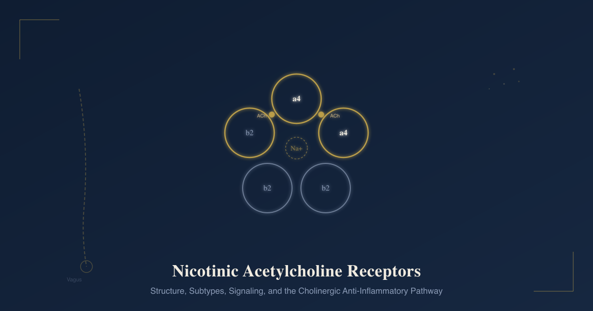
Subunit Architecture And The Pentameric Pore
Every functional nAChR is a pentamer: five homologous subunits arranged symmetrically around a central water-filled ion-conducting pore. R
The total molecular weight is approximately 290 kDa.
There are 17 distinct nAChR subunit genes in vertebrates: α1-α10, β1-β4, γ, δ, and ε.
Each subunit has the same basic domain architecture:
Large extracellular N-terminal domain (ECD):
Contains the ligand-binding domain with six binding loops (Loops A-C on α subunits, Loops D-F on the complementary subunit).
Contains the signature cysteine loop.
Contains the main immunogenic region (MIR) on α1 subunits, the primary target of autoantibodies in myasthenia gravis.
Four transmembrane helices (TM1-TM4):
TM2 lines the ion-conducting pore and is the most critical for channel function.
Mutations in TM2 alter ion selectivity and channel kinetics.
The leucine residues at position 9' in TM2 form the main hydrophobic gate that closes the channel.
Large intracellular domain (ICD) between TM3 and TM4:
The most variable region between subunits.
Contains phosphorylation sites that regulate channel trafficking, assembly, and function.
Contains sites for binding intracellular scaffold proteins.
Short extracellular C-terminal domain.
Alpha vs. non-alpha subunits:
Alpha subunits are defined by the presence of a vicinal cysteine pair (two adjacent cysteines, Cys191-Cys192 in the numbering based on Torpedo α1) near the entrance to TM1.
This cysteine pair participates in agonist binding and is unique to α subunits.
The agonist (ACh, nicotine) binds at the interface between an α subunit and its neighboring subunit.
The α subunit provides the principal binding face (Loops A, B, C).
The neighboring subunit provides the complementary binding face (Loops D, E, F).
In heteromeric receptors, the complementary face is a β subunit.
In homomeric α7 receptors, the complementary face is another α7 subunit.
Each pentamer has two agonist binding sites (one per α subunit, at the α-neighbor interface) and requires binding at both to activate.
Conformational States: Resting, Open, Desensitized
nAChRs are allosteric proteins that exist in multiple interconvertible conformational states: R
Resting state (closed, low agonist affinity):
The channel is closed.
The receptor has low affinity for agonist.
This is the baseline state in the absence of agonist.
Activated (open) state:
Agonist binds both sites, inducing conformational changes that propagate through the extracellular domain to the transmembrane helices.
The TM2 helices rotate, opening the hydrophobic gate.
The channel opens within milliseconds.
Na+ and K+ flow down their electrochemical gradients (predominantly Na+ influx).
Depolarization occurs.
Duration: milliseconds (for most heteromeric receptors) to tens of milliseconds (for α7).
Desensitized state (closed, high agonist affinity):
If agonist remains bound, the receptor undergoes a second conformational change into a non-conducting state.
The channel is closed despite agonist still being bound.
Crucially, the desensitized receptor has higher affinity for agonist than the resting state.
Recovery from desensitization requires agonist dissociation and is much slower than activation (milliseconds to seconds for rapid desensitization, hours to days for some long-term states).
This desensitization mechanism is central to understanding both nicotine addiction and nAChR pharmacology.
Nicotine's peculiarity is that it cannot be rapidly removed from the synapse by hydrolysis the way acetylcholine can.
Acetylcholinesterase rapidly degrades ACh, terminating the signal cleanly.
Nicotine persists.
Persistent agonist drives prolonged desensitization. R
Muscle-Type nAChRs: The Neuromuscular Junction
The neuromuscular junction (NMJ) is the synapse between a motor neuron axon terminal and a skeletal muscle fiber.
It is the site of the most intensively studied nAChR: the muscle-type nAChR.
Embryonic form: (α1)₂β1γδ
Adult form (from birth onward in most muscles): (α1)₂β1δε
The γ subunit is replaced by ε during development, which increases single-channel conductance and shortens mean open time, improving the speed and fidelity of neuromuscular transmission in adult muscle.
The two ACh binding sites are at the α1-δ and α1-ε (or α1-γ in embryonic) interfaces.
The NMJ transmission process:
- Motor neuron action potential arrives at the presynaptic terminal.
- Voltage-gated Ca2+ channels open, Ca2+ influx triggers vesicle fusion.
- ACh is released into the synaptic cleft.
- ACh diffuses to postsynaptic muscle nAChRs (densely clustered via rapsyn and agrin scaffolding).
- ACh binding opens nAChR ion channels.
- Na+ influx depolarizes the endplate (endplate potential, EPP).
- EPP reaches threshold and triggers a muscle action potential.
- ACh is rapidly hydrolyzed by acetylcholinesterase (AChE) in the synaptic cleft.
The NMJ has a substantial safety factor: EPP amplitude is normally 3-4 times the threshold needed for a muscle action potential, providing a buffer against partial transmission failure.
nAChR clustering at the NMJ is maintained by the agrin-MuSK-rapsyn signaling pathway:
- Agrin (secreted by the motor neuron) binds LRP4
- LRP4 activates MuSK (muscle-specific kinase)
- MuSK phosphorylates and activates rapsyn
- Rapsyn directly crosslinks nAChRs into dense postsynaptic clusters
Failure of any step in this pathway causes NMJ dysfunction. R
Neuronal nAChR Subtypes
The neuronal nAChR family uses a different subunit set: α2-α7, α9, α10 (α8 exists only in birds) and β2-β4.
These form an enormous diversity of possible heteromeric combinations.
Because each subunit has sidedness and the two binding sites have different pharmacological properties depending on which subunits form each interface, the potential for pharmacological specificity across subtypes is high. R
The two most common and most important neuronal subtypes are:
- α4β2 heteromeric receptors: high-affinity nicotine receptors, the dominant type in the brain
- α7 homomeric receptors: fast-activating, rapidly desensitizing, high calcium permeability, dominant in the immune system
The asterisk notation used in the literature (e.g., α4β2*, α7*) indicates that the receptor contains the named subunits but may contain additional subunits not specified.
α4β2: The High-Affinity Brain Receptor
α4β2 nAChRs are the most abundant nicotinic receptors in the mammalian brain and carry the highest affinity for nicotine (Ki ~1 nM for nicotine binding) among brain nAChRs.
They are heteromeric, requiring both α4 and β2 subunits to form functional receptors.
Stoichiometry diversity: α4β2 receptors can assemble as either (α4)₂(β2)₃ (high sensitivity, lower conductance, more easily desensitized) or (α4)₃(β2)₂ (lower sensitivity, higher conductance, less desensitization).
The ratio of these stoichiometries differs by brain region and affects pharmacological responses. R
α5 as an accessory subunit: α5 does not contribute to the agonist binding site but can incorporate as a fifth subunit into α4β2 complexes.
α5 inclusion enhances receptor assembly, reduces upregulation by nicotine, and facilitates channel closure.
The α5 subunit gene (CHRNA5) is in a chromosomal cluster at 15q24-25 with CHRNA3 and CHRNB4 that is a major genetic risk locus for nicotine dependence, lung cancer, and COPD. R
α4β2 distribution: Broad throughout the brain, particularly dense in:
- Ventral tegmental area (VTA) and substantia nigra: dopamine neuron cell bodies
- Nucleus accumbens (NAc): dopamine terminal regions
- Cortex and thalamus: attention and working memory circuits
- Hippocampus: memory consolidation
- Basal ganglia: motor control
What α4β2 does:
On dopamine neuron cell bodies in the VTA, α4β2 activation increases firing rate and burst firing, triggering dopamine release in the nucleus accumbens.
On GABAergic interneurons in the VTA, α4β2 activation drives inhibitory tone onto dopamine neurons.
The net effect of nicotine on dopamine output depends on the balance between direct excitation of dopamine neurons and indirect GABA-mediated inhibition, with desensitization of GABAergic α4β2 receptors eventually tipping the balance toward enhanced dopamine output (discussed below).
α7: The Homomeric Outlier
α7 nAChRs are unusual in multiple ways that distinguish them from every other nAChR subtype: R
Homomeric assembly: Five identical α7 subunits form the receptor.
Each α7-α7 interface contributes one binding site, giving five potential binding sites per pentamer (though functional studies suggest two are sufficient for activation).
High calcium permeability: α7 nAChRs are the most calcium-permeable of all nAChR subtypes.
The Ca2+ to Na+ permeability ratio (PCa/PNa) is approximately 10 for α7, compared to approximately 1.5 for most other subtypes.
This high Ca2+ permeability makes α7 activation functionally resemble NMDA receptor activation in some respects: it triggers Ca2+-dependent intracellular signaling cascades rather than just depolarization.
Extremely rapid desensitization: α7 receptors desensitize within milliseconds of agonist binding.
This is far faster than other nAChR subtypes.
It means α7 activation in response to brief ACh transients can generate a significant Ca2+ signal without the channel staying open long enough to depolarize significantly.
α-bungarotoxin sensitivity: α7 receptors are blocked by α-bungarotoxin (the snake toxin that historically defined the muscle nAChR) while other neuronal subtypes are resistant.
This pharmacological distinction was historically confusing (brain tissue that doesn't bind nicotine with high affinity but does bind α-bungarotoxin) before α7 was cloned and characterized.
Ionotropic and metabotropic signaling: α7 signals not only through its ion channel function but also through non-ionotropic (metabotropic-like) signaling pathways.
These include JAK2/STAT3 signaling, PI3K/Akt signaling, and interactions with the tumor necrosis factor receptor-associated factors (TRAFs), all through protein-protein interactions with the intracellular domain.
This metabotropic signaling from α7 is particularly important for its anti-inflammatory functions.
α7 distribution:
- CNS: hippocampus, cortex, ventral tegmental area, nucleus accumbens (presynaptic terminals on glutamatergic axons)
- Peripheral nervous system: sensory and autonomic ganglia
- Immune system: macrophages, lymphocytes, dendritic cells, microglia
- Non-neuronal: lung epithelium, vascular endothelium, gut epithelium, keratinocytes
The dual presence in the nervous system and immune system makes α7 the primary molecular mediator of the cholinergic anti-inflammatory pathway (discussed below).
Other Neuronal Subtypes
α3β4: The dominant subtype in autonomic ganglia (both sympathetic and parasympathetic).
α3β4 receptors mediate ganglionic transmission throughout the autonomic nervous system.
Mecamylamine, a classic nAChR antagonist used historically for hypertension, blocks α3β4 among other subtypes.
α3β4 is also the receptor that mediates the aversive (nausea-inducing) effects of nicotine at high doses, located in the medial habenula-interpeduncular nucleus (MHb-IPN) pathway.
Habenular α5-containing α3β4 receptors are a critical brake on nicotine consumption: α5 signals aversion, limiting how much nicotine is self-administered.
CHRNA5 polymorphisms that reduce aversive signaling through this pathway increase vulnerability to heavy smoking.
α6β2 (with β3 and α4 as accessory subunits): Restricted to catecholaminergic neurons, particularly in the mesolimbic dopamine pathway.
α6β2* receptors are on dopamine terminals in the nucleus accumbens and modulate phasic dopamine release.
α6 knockout mice show reduced nicotine-induced dopamine release.
α6β2* receptors have even higher nicotine sensitivity than α4β2 receptors in some striatal populations.
α9 and α9α10: Found in cochlear hair cells (critical for lateral efferent suppression of auditory transduction), vestibular hair cells, and immune cells including lymphocytes.
α9-containing receptors mediate neuropathic pain modulation through immune and sensory cell mechanisms. R
Non-Neuronal nAChRs
The discovery that nAChR subunit genes are expressed in non-neuronal tissues transformed the field's understanding of cholinergic signaling. R
Non-neuronal cholinergic systems operate independently of classical synaptic transmission, through autocrine and paracrine ACh release from non-neuronal cells.
Keratinocytes: Express α3, α7, α9, α10. ACh from keratinocytes (non-neuronal keratinocyte-derived ACh) activates nAChRs to regulate adhesion, differentiation, migration, and apoptosis.
Airway epithelium: α7 and α4β2 expression. Relevant to nicotine's effects on airway inflammation and to nicotine-related lung pathology.
Macrophages and immune cells: α7 primarily; critical for the cholinergic anti-inflammatory pathway.
Gut: Enteric neurons express multiple nAChR subtypes regulating gut motility. Gut epithelial cells express α7 relevant to gut inflammation.
Vascular endothelium: α7 expression relevant to vascular inflammation and endothelial function.
Pancreatic beta cells: nAChR expression linked to insulin secretion modulation.
Nicotine Pharmacology And Addiction
Understanding nicotine's mechanism requires starting from the normal physiology of acetylcholine at nAChRs and recognizing where nicotine deviates.
Normal ACh Signaling
ACh is released in brief, high-concentration pulses from presynaptic terminals.
Acetylcholinesterase in the synapse rapidly hydrolyzes ACh within milliseconds.
The result: brief receptor activation, clean termination, and recovery from desensitization before the next pulse.
Nicotine's Deviation
Nicotine is not hydrolyzed by acetylcholinesterase.
It persists in the synapse after inhalation, maintaining receptor occupancy.
Brain nicotine concentrations from smoking reach 20-100 nM, enough to occupy high-affinity α4β2 receptors. R
Sustained low-level nicotine causes a complex temporal sequence of receptor activation followed by desensitization:
Phase 1 (minutes after smoking):
Nicotine activates α4β2* nAChRs on VTA dopamine neurons and on presynaptic glutamatergic terminals projecting to the VTA.
Both activations increase dopamine neuron firing.
α7* nAChRs on glutamatergic presynaptic terminals enhance glutamate release onto VTA dopamine neurons, increasing their burst firing.
Dopamine floods the nucleus accumbens, driving reward and reinforcement.
Phase 2 (continuing minutes):
With sustained nicotine, α4β2* nAChRs on GABAergic interneurons in the VTA desensitize.
These GABAergic nAChRs have lower desensitization resistance than dopaminergic nAChRs.
As GABAergic tone drops (due to desensitized α4β2 on GABA neurons), the inhibitory brake on dopamine neurons lifts.
This disinhibition further enhances dopamine neuron activity through a second, delayed wave of excitation.
α4β2 desensitization on dopamine neurons themselves occurs more slowly.
The net effect over a cigarette: a burst of dopamine release followed by a prolonged period of enhanced dopamine excitability from disinhibition, with progressively declining direct activation as receptors desensitize. R
Upregulation: The Paradox
Chronic nicotine exposure causes a paradoxical increase in the number of α4β2* binding sites in the brain.
This upregulation is not at the mRNA level.
It occurs post-translationally: nicotine-stabilized desensitized receptors accumulate in intracellular compartments, reducing receptor degradation, resulting in a net increase in receptor protein.
The result is a larger total pool of nAChRs, most of which are in the desensitized state during active smoking.
This upregulation is maintained as long as nicotine is present.
When smoking ceases:
- Nicotine clears over hours
- Upregulated α4β2 receptors recover from desensitization
- The enlarged receptor pool is now activated by normal ACh
- ACh-driven nAChR activity surges
- This surge contributes to the anxiety, irritability, craving, and cognitive deficits of nicotine withdrawal R
Brain imaging (PET) studies confirm that α4β2 receptor density positively correlates with withdrawal difficulty in smokers.
The Cholinergic Anti-Inflammatory Pathway
This is arguably the most important nAChR function outside the nervous system, and it is substantially underappreciated in clinical medicine. R
The discovery:
In 1998-2003, Kevin Tracey's lab at the Feinstein Institute demonstrated that the efferent vagus nerve could suppress systemic inflammatory responses to endotoxin.
They showed that vagotomy dramatically increased TNF-alpha production in response to LPS, while electrical vagal nerve stimulation suppressed it.
The active molecule was acetylcholine.
The critical receptor on macrophages was α7 nAChR.
The pathway was named the cholinergic anti-inflammatory pathway (CAP). R
The anatomy:
The vagus nerve carries both sensory (afferent) and motor (efferent) fibers.
Afferent vagal fibers detect pro-inflammatory cytokines in peripheral tissues and relay this signal to the brainstem.
The brainstem integrates this with other signals and activates efferent vagal outflow.
The efferent vagus does not directly innervate macrophages in the spleen.
Instead, vagal efferent signals activate the celiac/splenic nerve, which is sympathetic.
The splenic nerve terminates on a specialized population of T cells in the spleen that synthesize and release ACh. R
ACh-synthesizing splenic T cells release ACh onto α7 nAChR-expressing macrophages.
The molecular mechanism:
ACh binding to macrophage α7 nAChRs activates the JAK2/STAT3 pathway.
Phosphorylated STAT3 translocates to the nucleus and blocks NF-kB p65 transcription.
Simultaneously, α7 activation prevents IκB phosphorylation and degradation, keeping NF-kB in its inhibited form.
The result: suppressed production of TNF-alpha, IL-1β, IL-6, and IL-18. R
Critically, IL-10 (the anti-inflammatory cytokine) is not suppressed by this pathway.
The CAP is therefore selectively anti-inflammatory: it reduces the pro-inflammatory cytokine storm while leaving anti-inflammatory resolution signals intact.
Disease relevance of the CAP:
The CAP is protective in experimental models of sepsis, inflammatory bowel disease, rheumatoid arthritis, kidney injury, stroke, and metabolic syndrome.
Vagal nerve stimulation (VNS), which activates the CAP, has shown clinical benefit in rheumatoid arthritis, Crohn's disease, and IBD in human clinical trials.
Implantable VNS devices are in active clinical development specifically for this anti-inflammatory mechanism.
The CAP may partly explain why inflammatory conditions (IBD, rheumatoid arthritis, Crohn's disease) show paradoxically reduced prevalence in smokers compared to non-smokers, a well-documented but ethically complicated epidemiological observation.
Nicotine activates macrophage α7 nAChRs directly, mimicking the afferent arm of the CAP. R
This anti-inflammatory mechanism via α7 is therapeutically interesting because it suggests that selective α7 agonists could activate the CAP without the addictive liability of nicotine at α4β2 receptors.
Disease States And nAChR Dysfunction
Myasthenia Gravis
Myasthenia gravis (MG) is an autoimmune disorder of the NMJ caused primarily by autoantibodies against muscle nAChRs. R
The autoantibodies are directed primarily at the main immunogenic region (MIR) of the α1 subunit (residues 67-76).
They cause disease through three mechanisms:
- Direct blockade of ACh binding sites
- Crosslinking and accelerated internalization and degradation of nAChRs (antigenic modulation), reducing receptor density
- Complement activation at the endplate, damaging the postsynaptic membrane
The reduced receptor density and damaged membrane reduce EPP amplitude below the threshold required for muscle action potential generation, causing the characteristic fatigable weakness.
Clinical features: Fluctuating muscle weakness that worsens with activity and improves with rest.
Initial extraocular involvement causes ptosis and diplopia.
Progression to bulbar (dysphagia, dysarthria) and limb involvement.
MuSK antibody MG (10-15% of cases): Autoantibodies against MuSK rather than nAChR directly; disrupts rapsyn-mediated nAChR clustering; different distribution of weakness (more bulbar and respiratory).
Treatment: Acetylcholinesterase inhibitors (pyridostigmine) increase synaptic ACh dwell time, compensating for reduced receptor density. Immunosuppression (steroids, azathioprine, mycophenolate). Thymectomy. Biological agents targeting B cells or complement for refractory cases.
Congenital Myasthenic Syndromes
Congenital myasthenic syndromes (CMS) are inherited disorders of NMJ function caused by mutations in genes encoding NMJ proteins, including nAChR subunit genes themselves (α1, β1, δ, ε). R
Slow-channel CMS: Gain-of-function mutations in nAChR subunits that prolong channel opening time.
The prolonged Ca2+ influx causes endplate myopathy from Ca2+ overload.
Treatment: quinidine or fluoxetine to reduce channel open time.
Fast-channel CMS: Loss-of-function mutations that shorten or reduce channel opening.
Treatment: AChE inhibitors and 3,4-diaminopyridine (increases ACh release).
AChR deficiency CMS: Loss-of-expression mutations.
Treatment: AChE inhibitors.
Alzheimer's Disease
Cortical and hippocampal nAChR expression, particularly α7 and α4β2, is substantially reduced in Alzheimer's disease.
α7 nAChRs are lost early in the disease process.
α7 nAChRs interact with amyloid beta oligomers, with Aβ42 acting as a partial agonist or antagonist at α7 depending on concentration.
Cholinergic neuron degeneration (the basis for the cholinergic hypothesis of Alzheimer's) depletes the ACh that normally stimulates nAChRs throughout the cortex.
AChE inhibitors (donepezil, rivastigmine, galantamine) used in Alzheimer's treatment work partly through preserving nAChR activation.
Selective α7 agonists are in clinical development for Alzheimer's cognitive symptoms. R
Schizophrenia
α7 nAChR expression and function is reduced in schizophrenia.
The gene encoding α7 (CHRNA7 on chromosome 15q14) has been linked to schizophrenia endophenotypes.
CHRNA7 promoter polymorphisms that reduce α7 expression are associated with schizophrenia.
Reduced α7 function impairs sensory gating (the brain's ability to suppress repetitive stimuli), a core defect in schizophrenia.
This likely explains the extraordinarily high smoking rates in schizophrenia (70-90%): patients self-medicate with nicotine to activate their deficient α7 receptors and temporarily restore sensory gating.
α7 selective agonists and positive allosteric modulators (PAMs) are in clinical development for schizophrenia. R
Parkinson's Disease
nAChR expression in the substantia nigra and striatum is reduced in Parkinson's disease in parallel with dopaminergic neuron loss.
α6β2* nAChRs, which are concentrated on dopaminergic terminals, are among the earliest casualties.
Epidemiological studies consistently find lower Parkinson's disease incidence in smokers, suggesting some neuroprotective effect of nicotine, though confounders make this difficult to interpret.
NMN-linked α4β2* receptor activation may support dopaminergic neuron survival through BDNF and neuroprotective signaling.
Epilepsy
Autosomal dominant nocturnal frontal lobe epilepsy (ADNFLE) is caused by mutations in CHRNA4 (α4) or CHRNB2 (β2) genes.
Mutations that cause gain-of-function (increased sensitivity or reduced desensitization) in α4β2 receptors lead to excessive cholinergic excitation during sleep, triggering seizures.
CHRNA2 is a third candidate gene for ADNFLE.
Pharmacology: Agonists, Antagonists, And Modulators
Endogenous Agonist
Acetylcholine (ACh): binds all nAChRs, rapidly hydrolyzed.
Choline: a partial agonist selective for α7 (major breakdown product of ACh after hydrolysis).
Pharmacological Agonists
Nicotine: partial agonist at most nAChR subtypes; more potent at α4β2 than α7; not hydrolyzed by AChE; causes desensitization under sustained exposure.
Epibatidine: derived from Ecuadorian poison dart frog skin; extremely potent full agonist at nAChRs; analgesic but toxic at therapeutic doses.
GTS-21 (DMXBA): partial agonist selective for α7; used in anti-inflammatory and cognitive research.
PNU-282987: full agonist selective for α7; used as a research tool and in CAP research.
Cytisine: partial agonist at α4β2; used as a smoking cessation agent in Eastern Europe for decades; recently approved as Cytisinicline (Cytisine, 1.5 mg) in some markets; weaker partial agonist than varenicline.
Partial Agonists With Therapeutic Application
Varenicline (Chantix/Champix):
Partial agonist at α4β2* nAChRs (Ki ~1 nM), full agonist at α7 nAChRs. R
Mechanism of smoking cessation action: as a partial agonist at α4β2, it provides enough receptor stimulation to reduce craving (more than zero ACh activity), while blocking nicotine's higher-efficacy stimulation when a cigarette is smoked (competitive inhibition).
The result: reduced dopamine release from smoking (because nicotine cannot displace varenicline fully), reduced reward, reduced reinforcement.
Varenicline also desensitizes nAChRs to some degree.
It achieves significantly better quit rates than bupropion or nicotine replacement therapy in head-to-head trials.
Side effects include nausea (α7 activation in the gut/brainstem), vivid dreams, and historically debated neuropsychiatric effects (current evidence suggests risk is low).
Antagonists
α-Bungarotoxin (αBTX):
Snake toxin from Bungarus multicinctus; irreversible competitive antagonist at α1 (muscle) and α7 (neuronal) nAChRs; does not block most other neuronal subtypes.
Essential research tool for isolating α7 responses and for purifying muscle nAChRs.
Mecamylamine:
Non-competitive (channel-blocking) antagonist; crosses the blood-brain barrier; blocks all neuronal nAChRs nonselectively.
Historically used as an antihypertensive (via ganglionic blockade of sympathetic ganglia α3β4 receptors).
Has antidepressant properties in mice, likely through β2 nAChR blockade.
Being studied as an adjunct to nicotine replacement for smoking cessation, because nAChR desensitization/blockade may reduce the aversive aspects of quitting.
Dihydro-β-erythroidine (DHβE):
Competitive antagonist; selective for α4β2 over α7; does not cross the BBB well; research tool.
α-Conotoxin MII:
Peptide toxin from cone snails; selective for α6β2-containing receptors; research tool for isolating α6β2 contributions to dopamine release.
Tubocurarine (d-TC) and pancuronium:
Non-depolarizing neuromuscular blocking agents; competitive antagonists at NMJ muscle nAChRs; used for surgical muscle relaxation.
Succinylcholine:
Depolarizing NMJ blocker; acts as a non-hydrolyzed agonist at muscle nAChRs, producing persistent depolarization and a flaccid paralysis that lasts until succinylcholine is metabolized by pseudocholinesterase.
Positive Allosteric Modulators (PAMs)
PAMs bind outside the agonist binding site and enhance receptor responsiveness without activating the channel themselves.
Type I PAMs: Increase peak current amplitude without slowing desensitization.
Type II PAMs: Increase peak current and slow desensitization, effectively reactivating desensitized receptors.
Type II PAMs at α7 (such as PNU-120596) produce dramatically larger and more prolonged α7 currents, potentially amplifying the anti-inflammatory effects of the CAP.
Type II α7 PAMs are in preclinical development for schizophrenia, Alzheimer's, and inflammatory diseases.
What To Stay Away From
- Assuming nicotinic and muscarinic receptors are interchangeable: they are both activated by ACh and share the "cholinergic" designation, but they are structurally unrelated and functionally opposite in many contexts; muscarinic receptors are GPCRs (Gq, Gi, Gs-coupled depending on subtype); nicotinic receptors are ligand-gated ion channels; drugs that act on one class do not act on the other, and many clinical drugs are selective for one family (atropine blocks muscarinic, mecamylamine blocks nicotinic)
- Long-term high-dose nicotine without recognizing α4β2 upregulation and desensitization dynamics: the counter-intuitive consequence of chronic nicotine is that it creates a state of receptor upregulation combined with chronic desensitization, and abrupt cessation unmasks the upregulated receptor pool responding vigorously to endogenous ACh, driving withdrawal; tapering is pharmacologically rational
- Misinterpreting epidemiological protection of smoking against Parkinson's and UC without mechanistic context: the nAChR-mediated mechanisms (dopaminergic neuroprotection via α6β2 in Parkinson's, α7-CAP activation in UC) are real, but cigarette smoke contains hundreds of additional toxic compounds; the public health calculus is unambiguous; this is biology of interest for α7 agonist drug development, not a rationale for smoking
- G6PD-deficient patients and high-dose nicotine or galantamine: some individuals with G6PD deficiency metabolize cholinergic agents differently and may have altered responses to AChE inhibitors
- AChE inhibitor use without recognizing that it amplifies all cholinergic actions: donepezil, rivastigmine, and galantamine increase synaptic ACh, which activates both nicotinic and muscarinic receptors; muscarinic side effects (bradycardia, increased secretions, GI cramps, nausea) are common and reflect M2/M3 receptor activation; these are distinct from nAChR effects
Mechanisms Of Action
Simple:
- Nicotinic receptors are like gated doors in the cell membrane: when acetylcholine (or nicotine) binds to two spots on the outside of the door, it swings open for a fraction of a second, letting sodium rush in and briefly exciting the cell; when the door stays open too long because nicotine keeps it jammed, the cell adapts by closing the door even while nicotine is still there, which is called desensitization.
- When you smoke a cigarette, nicotine activates the α4β2 receptors on dopamine neurons in the brain's reward center, triggering a dopamine pulse that the brain learns to associate with the cigarette; simultaneously, nicotine desensitizes the same type of receptor on inhibitory neurons nearby, which eventually removes the brake on dopamine neurons and prolongs the high.
- Over weeks of smoking, the brain compensates for being chronically jammed by growing more α4β2 receptors; when you quit, all those extra receptors suddenly start responding to normal acetylcholine and flood the reward system, which is why withdrawal feels so uncomfortable.
- The vagus nerve uses α7 receptors on macrophages as an anti-inflammatory off switch: when the vagus fires, it eventually triggers acetylcholine release in the spleen, which lands on α7 receptors on immune cells and tells them to stop making inflammatory cytokines like TNF and IL-1; this is why nerve stimulation implants are being tested for Crohn's disease and rheumatoid arthritis.
- Myasthenia gravis happens when the immune system makes antibodies against the muscle version of the nicotinic receptor at the neuromuscular junction, accelerating its destruction; the resulting shortage of receptors means that nerve signals often fail to trigger muscle contraction, producing the characteristic weakness that gets worse with use and better with rest.
Advanced:
- Conformational state transitions and the basis for desensitization: The nAChR is an allosteric protein with a free energy landscape that includes at least three major stable states. The resting closed state (low agonist affinity, Kd ~1 µM for ACh) transitions upon agonist binding to the open state (Kd ~0.01 µM, high affinity) through a concerted conformational change that involves outward rotation of the transmembrane helices, particularly TM2, expanding the hydrophobic gate. The open state is kinetically unstable and transitions rapidly to the desensitized state (also high affinity, ~0.01 µM, but channel closed). The thermodynamic link between ligand affinity and channel state explains why high concentrations of agonist preferentially accumulate the desensitized state: the high-affinity desensitized receptor captures and traps the ligand. This kinetic mechanism means that nicotine (which cannot be cleared by AChE) drives the receptor population progressively into the desensitized state over minutes, while ACh pulses produce brief open-state transients that readily recover. R
- The α4β2 desensitization-disinhibition mechanism in nicotine addiction: VTA GABA interneurons express α4β2* nAChRs at higher density and with lower desensitization resistance than neighboring dopamine neurons. Nicotine initially activates both populations. Within minutes, GABAergic α4β2* desensitize, reducing inhibitory tone onto dopamine neurons; dopaminergic α4β2* remain partially active longer. This temporal dissociation creates a "disinhibition window" during which dopamine neurons fire more because their GABA brake is off while their own excitatory drive remains partially intact. The signal-to-noise improvement in dopamine release (less tonic background dopamine "noise" from desensitized GABAergic inputs, preserved phasic burst firing from dopaminergic inputs) may encode nicotine's association with environmental cues particularly efficiently, contributing to the robustness of nicotine-associated conditioned reinforcement. R
- α7 metabotropic signaling and the CAP molecular mechanism: The classic view of nAChRs as purely ionotropic (generating currents) is incomplete for α7. Macrophage α7 nAChRs signal through at least two ion-independent pathways. First, α7 recruits JAK2 at the intracellular domain, activating the JAK2/STAT3 pathway; phosphorylated STAT3 forms a complex with NF-κB p65 that blocks NF-κB-driven transcription of TNF, IL-1β, IL-6, and IL-18 without affecting IL-10 synthesis. Second, α7 activation prevents IκB kinase (IKK) phosphorylation, keeping IκB bound to NF-κB and preventing its nuclear translocation. Both mechanisms converge on NF-κB inhibition. The high Ca2+ permeability of α7 also contributes: Ca2+ influx can activate Ca2+-dependent kinases that phosphorylate and inactivate inflammatory signaling components. Type II PAMs that prevent α7 desensitization amplify all three pathways and produce dramatically enhanced anti-inflammatory effects in preclinical models. R
More Research
- Nicotinic receptor genetics are directly clinically relevant: the CHRNA5/A3/B4 gene cluster at 15q24-25 is one of the strongest genetic risk loci for nicotine dependence, lung cancer, COPD, and peripheral arterial disease; CHRNA5 polymorphisms (particularly the D398N variant, rs16969968) reduce aversive signaling through habenular α5-containing receptors, increasing vulnerability to heavy smoking by reducing the natural limit on nicotine intake. R
- Vagus nerve stimulation (VNS) works through the cholinergic anti-inflammatory pathway via α7 nAChR on macrophages; clinical trials for rheumatoid arthritis and IBD have shown anti-inflammatory benefit from implantable VNS devices; this represents one of the first therapeutic applications of neuroimmune circuitry in human disease; selective α7 agonists (GTS-21 and others) are being developed to pharmacologically activate the CAP without requiring nerve stimulation. R
- α7 nAChR dysfunction is central to multiple psychiatric diseases including schizophrenia (reduced α7 expression causes sensory gating deficit, driving nicotine self-medication), Alzheimer's disease (α7 is lost early, interacts with amyloid beta), and depression (α7 CAP activation has antidepressant effects via neuroinflammation reduction); the smoking rates in schizophrenia (70-90%) are substantially explained by this nAChR deficit rather than by non-specific psychiatric risk factors. R
- Varenicline as a smoking cessation agent works through partial agonism at α4β2 nAChRs: it provides enough stimulation to reduce craving while blocking the reinforcement that comes from smoking a cigarette on top of it; PET studies show that therapeutic varenicline doses occupy approximately 90% of α4β2 receptors in the brain; the drug also activates α7 fully, contributing to cognitive effects but also to nausea. R
- Congenital myasthenic syndromes are a window into the functional importance of every NMJ protein: mutations in nAChR subunits (α1, β1, δ, ε), AChE anchoring protein COLQ, the clustering scaffold rapsyn, the receptor tyrosine kinase MuSK, and downstream signaling proteins all cause NMJ transmission failure with characteristic electrophysiological and clinical signatures; genetic diagnosis changes treatment because slow-channel CMS (treated with quinidine or fluoxetine) is worsened by AChE inhibitors that would treat fast-channel CMS. R
- The epidemiological association between smoking and reduced risk of Parkinson's disease and ulcerative colitis is real and likely involves α7-CAP activation (UC) and α6β2/α4β2 dopaminergic neuroprotection (Parkinson's); these observations are driving selective α7 agonist and PAM development for inflammatory and neurodegenerative indications as a way to access these protective mechanisms without tobacco's carcinogenic, cardiovascular, and addictive effects.
Jacob Gordon
INHC, FMT-C
Board Certified Health Coach
I spent years battling unexplained chronic illness before discovering biohacking, epigenetics, and functional medicine. Now I share that research at MyBioHack to help others find their own answers.
Book a ConsultationRelated Protocols & Supplements
Deep-dive chapters and recommended supplements for this topic
Quercetin
500mg 2x/day
Vitamin D3 + K2
5000 IU + 200mcg/day
DAO Enzyme
1 cap before meals