Mast Cells (Part 3): The Mast Cell-Glia Interaction In Chronic Inflammation
By Jacob Gordon, INHC, FMT-CThis article contains affiliate links. As an Amazon Associate, MyBioHack earns from qualifying purchases at no extra cost to you. We only link products we research and stand behind.

This is a multi-part series on mast cells:
Mast Cells (Part 1): Stabilize Mast Cells And Remove Brain Fog
Mast Cells (Part 3): The Mast Cell-Glia Interaction In Chronic Inflammation
In this post we will focus on the mast cell-glia (microglia, astrocyte, oligodendrocyte) interaction with our body and how they contribute to chronic inflammatory diseases such as CIRS and SALI.
Basics
The Central Nervous System (CNS) can communicate to the immune system and mediate multiple inflammatory responses.
While inflammation per se may not cause disease, it contributes importantly to disease pathogenesis across both the peripheral and central nervous systems: R
Alzheimer Disease
Amyotrophic Lateral Sclerosis
Autism Spectrum Disorder
Depression
Fibromyalgia
Ischemia
Motor Neuron Disease
Multiple Sclerosis
Neuropathic Pain
Parkinson Disease
Traumatic Brain Injury
The core problem with inflammation is not how often it starts, but how often it fails to subside. R
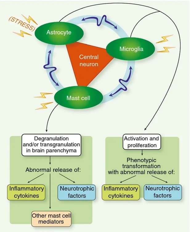
Microglia (the brain's main immune guardians) and mast cells, along with astrocytes (and possibly even oligodendrocytes) are the three biggest players in chronic inflammation.
They all play a key role by regulating our responses to infection, inflammation, and reactions to stress or trauma.
Just looking at these 3 parts individually does not provide a whole picture of SALI but does highly represent the chronic inflammatory response.
Glia
Microglia provide “immunosurveliance” of the brain, constantly surveying their environment in preparation for insult or injury. R
When they are activated, they eat up cellular debris and then present antigens to T cells and release cytokines/chemokines. R
These processes are normal and used to keep homeostasis in the brain, such as: R
Engulf synaptic material
Initiate synaptic pruning
Regulate cell death
Regulate neurogenesis
M1 Vs M2 State Microglia
Microglia play a role in plasticity and will either be in a M1 (classic/pro-inflammatory) or M2 (alternative polarization/neuroprotective) state. R
Astrocytes

Astrocytes act like “brain glue” in the brain and regulate Blood-Brain-Barrier (BBB) integrity, axonal growth, and myelination. R
Oligodendrocytes
Oligodendrocytes help in myelin production as well as providing trophic support for axons and support for ATP production (via monocarboxylate transporter 1). R
Mast Cells
Mast cells play a multitude of functions and generally act as environmental sensors. R
When activated they can release a numerous amount “packaged” molecules such as numerous vasoactive, neurosensitizing and pro-inflammatory mediators, which include biogenic amines (histamine, serotonin), cytokines, proteolytic enzymes (e.g., chymase, tryptase, acid hydrolases, among others), lipid metabolites (prostaglandin D2, leukotriene C4, platelet-activating factor), ATP, neuropeptides, nerve growth factor (NGF), vascular endothelial growth factor (VEGF) and nitric oxide. R R
Mast cells can also induce T cell activation, proliferation, and cytokine secretion. R
How Mast Cells And Glia Create Strong Bonds

The more inflammation happens, the more that mast cells and glia communicate and create stronger bonds/crosstalk.
This mast cell-glia/glia-glia crosstalk can be mediated via: R
Antigen-presenting cells (CD40/CD40L/RhoGTPase/MAPK/NF-kb/STAT1) R
Central sensitization (BDNF hypersensitivity/ATP/P2/P2X4/Ca2+/NF-κB/PAR2/TNF-α/IL-6/IL-33/IL-13/CCL2) R
PAMPs (TLR2/TLR4/CCL5/RANTES/IL-6/CCL5) R
How Neuroinflammation Causes Inflammaging
Aging is associated with elevated levels of circulating cytokines and pro-inflammatory markers, and age-related changes in the immune system often referred to as “immunosenescence” or “inflammoaging”. R R
For example, infections have shown to cause inflammoaging of the hippocampus, a possible pathology of Alzheimer’s. R
Senescent (old, but undead) microglia are present in aging - they are primed to activation, but resistant to regulation. R R R
These “primed” kind of glia are extra sensitive to a secondary inflammatory stimulus, thus leading to an exaggerated inflammatory response, and may contribute to the inflammaging as seen in CIRS and SALI. R
For example, Lipopolysaccharides (LPS) cause endotoxemia (sepsis - showing a multipolar cytokine storm similar to CIRS), and LPS are able to prime the glia and mast cells. R
Having chronic levels of even low LPS can cause a persistent state of low-grade inflammation which is associated with innate immune “programming” or “memory” (ie altered BBB, cognitive dysfunction, pain, etc). R R
Top Ways To Stop Mast Cell-Glia Inflammation

Top Ways To Stop Mast Cell-Glia Inflammation:
Palmitoylethanolamide (PEA) R
Jacob Gordon
INHC, FMT-C
Board Certified Health Coach
I spent years battling unexplained chronic illness before discovering biohacking, epigenetics, and functional medicine. Now I share that research at MyBioHack to help others find their own answers.
Book a ConsultationRelated Protocols & Supplements
Deep-dive chapters and recommended supplements for this topic
Glutathione (Liposomal)
500mg 2x/day
Activated Charcoal
500mg away from food/meds
NAC
600mg 2x/day