HDAC Inhibition And Fear Extinction: The Epigenetic Key To Unlocking Traumatic Memory
By Jacob Gordon, INHC, FMT-CThis article contains affiliate links. As an Amazon Associate, MyBioHack earns from qualifying purchases at no extra cost to you. We only link products we research and stand behind.
The reason exposure therapy fails to produce lasting relief in so many people with post-traumatic stress disorder (PTSD) is not psychological resistance. It is a failure of consolidation, and the mechanism is epigenetic.
What Fear Extinction Actually Is
The most important thing to understand about fear extinction is what it is not.
It is not erasure.
The original fear memory is not deleted during extinction. It persists in the lateral nucleus of the basolateral amygdala (BLA), encoded in long-term potentiated synapses that were strengthened during the original conditioned stimulus (CS) / unconditioned stimulus (US) pairing. R
Fear extinction is new learning.
When a person or animal is repeatedly exposed to the conditioned stimulus without the aversive outcome, the brain forms a new inhibitory memory trace that actively competes with and suppresses the original fear memory. R
This distinction matters for everything that follows. Because the original memory is not erased, the extinction memory must continuously out-compete it. When the extinction memory weakens (as it does over time), when context changes, or when a new stressor occurs, the original fear memory resurfaces. This is why PTSD responds to exposure therapy initially and then relapses. R
The goal of treatment, then, is not to expose people to their fear and hope the original memory fades. The goal is to build an extinction memory that is robust enough to permanently dominate the fear memory in all contexts. That is where epigenetics enters.
The Neural Circuit: Amygdala, Infralimbic Cortex, Hippocampus
Three brain regions form the core fear-extinction circuit: the amygdala, the medial prefrontal cortex (mPFC), and the hippocampus.
The amygdala encodes and expresses fear. The lateral amygdala (LA) is where CS and US information converges and where the synaptic changes of fear conditioning are stored. The central amygdala (CeA) is the output nucleus: CeA activation drives downstream fear responses through the periaqueductal gray (PAG), producing freezing, autonomic arousal, and stress hormone release. R
Intercalated cells (ITC) are clusters of GABAergic inhibitory neurons positioned between the BLA and CeA. They function as a gate: when active, they suppress CeA output and therefore suppress fear expression. The infralimbic cortex activates these cells to shut the fear gate. R
The medial prefrontal cortex has two subregions with opposing functions: The prelimbic cortex (PL) drives fear expression. PL activation during retrieval of a conditioned stimulus sustains the fear response. R The infralimbic cortex (IL) drives extinction expression. IL neurons fire during retrieval of extinction memory, activating the ITC to suppress CeA output and silence the fear response. R
Stimulation of the vmPFC (ventromedial prefrontal cortex) to amygdala pathway facilitates extinction memory formation; silencing this pathway impairs extinction formation and reduces extinction-induced amygdala activity. R
The hippocampus provides contextual gating. The hippocampus tracks the context (the specific environment) in which extinction occurred. If the conditioned stimulus is encountered in a different context than where extinction training happened, the hippocampus communicates this mismatch to the BLA and PL, and the fear memory resurfaces rather than the extinction memory. R This is why exposure therapy conducted in a clinic does not always generalize to the real-world environments where the fear is triggered.
The practical implication: extinction memory is highly context-dependent. The original fear memory is context-independent. For extinction to prevail across environments, the extinction memory must be consolidated strongly enough at the circuit level to override contextual gating.
Why Extinction Fails: Relapse, Renewal, And Reinstatement
Even when exposure therapy produces apparent fear reduction, three relapse phenomena can restore the original fear response. R
Renewal occurs when the conditioned stimulus is encountered in a context different from where extinction training occurred. The hippocampus detects the context mismatch and reinstates hippocampal and PL inputs to the BLA, suppressing IL-driven extinction retrieval.
Reinstatement occurs when exposure to the original aversive stimulus (even in a context where extinction occurred) causes the fear memory to return. A single footshock after successful extinction is often sufficient to reinstate fear fully.
Spontaneous recovery is the time-dependent return of fear after extinction without any triggering event. Extinction memories fade faster than fear conditioning memories, and the fear memory gradually reasserts dominance over time.
All three forms of relapse reflect the same underlying fact: extinction is not erasure. The fear memory is always present, waiting for a shift in context, stress state, or time.
What would make extinction permanent? Consolidating the extinction memory at the synaptic and epigenetic level strongly enough that the extinction engram itself becomes as durable as the original fear engram. That is what histone deacetylase (HDAC) inhibition achieves. R
Epigenetic Gating Of Extinction Memory
Memory consolidation requires new protein synthesis and transcriptional changes in neurons. Not all experiences produce durable long-term memories. The signal strength, timing, and repetition of a learning event determine whether it crosses the threshold for long-term potentiation (LTP) and gene expression changes that encode lasting memory. R
Chromatin, the complex of DNA wound around histone proteins, regulates which genes can be transcribed. Histone acetyltransferases (HATs) add acetyl groups to histone tails, loosening the chromatin and allowing transcription factor access. Histone deacetylases (HDACs) remove those acetyl groups, tightening chromatin and repressing gene expression. R
Fear conditioning and fear extinction produce distinct and opposing patterns of histone acetylation in the mPFC and amygdala:
Fear conditioning increases histone H3 acetylation at BDNF (brain-derived neurotrophic factor) promoter regions in the lateral amygdala, upregulating BDNF expression to consolidate the fear trace. R
Fear extinction increases histone H4 acetylation specifically at BDNF exon IV promoter in the infralimbic cortex and hippocampus, driving the BDNF expression needed to consolidate the competing extinction memory. R
This differential acetylation of H3 (fear conditioning) versus H4 (fear extinction) at the same BDNF gene locus is not coincidental. It suggests that extinction is not simply the reversal of fear conditioning at the molecular level. It is a distinct learning event with its own epigenetic signature, and that signature can be strengthened or weakened by manipulating HDAC activity. R
When HDAC inhibitors are administered systemically, intrahippocampally, or directly into the infralimbic cortex before or after extinction training, extinction memory is enhanced: fear is lower during retrieval tests 24 hours later, and long-term fear inhibition is greater. R
The mechanism is not nonspecific memory enhancement across the board. Infusion of an HDAC inhibitor into the lateral amygdala immediately after fear conditioning can actually impair extinction by strengthening the fear memory itself. This region-specificity and timing-specificity is critical: HDAC inhibition is only beneficial for extinction when applied to the right brain regions at the right time, specifically the hippocampus and infralimbic cortex after extinction training. R
HDAC2 And The Neuroplasticity Window
Among the histone deacetylase isoforms, HDAC2 has emerged as the central regulator of the neuroplasticity window that determines whether extinction can consolidate. R
When a recent fear memory (formed within the past 24 hours) is retrieved, a cascade is triggered:
The retrieval event generates nitric oxide (NO) signaling in hippocampal neurons. NO causes S-nitrosylation of HDAC2, a post-translational modification on cysteine residues 262 and 274. R S-nitrosylation releases HDAC2 from chromatin at the promoters of neuroplasticity genes including c-Fos (an immediate early gene critical for neuronal activation). This release allows histone H3 acetylation to accumulate at these gene promoters. Histone acetylation opens chromatin at these loci, enabling transcription of plasticity genes, which increases synaptic flexibility, dendritic remodeling, and structural plasticity. R
This is the neuroplasticity window. It is a brief period (approximately 6 hours) following recent memory retrieval during which the synaptic substrate is labile and can be updated. Extinction training conducted during this window produces more durable fear inhibition than standard extinction conducted without retrieval. R
The critical problem for PTSD:
In remote memories (formed more than a month prior), retrieval does NOT trigger S-nitrosylation of HDAC2. HDAC2 stays bound to chromatin at neuroplasticity gene promoters. Histone acetylation fails to accumulate. The plasticity window does not open. Extinction training during this state consolidates poorly or not at all. R
This is the molecular explanation for why remote traumatic memories are so much harder to extinguish than recent ones, and why patients with long-standing PTSD respond less well to standard exposure therapy than those treated shortly after trauma.
Which HDACs Matter
Not all HDAC isoforms play the same role in extinction, and the specificity matters for understanding which inhibitors are therapeutically relevant.
Class I HDACs (HDAC1, HDAC2, HDAC3, HDAC8) are the primary regulators of fear extinction memory. R
HDAC1 and HDAC2 appear to be the primary negative regulators of extinction retention:
HDAC2 overexpression in the forebrain impairs context and cue-dependent fear learning. Conversely, forebrain-specific knockout of HDAC2 enhances both context fear and cued fear extinction. Modulation of HDAC1 expression alters context fear extinction, with HDAC1 overexpression actually enhancing extinction in some models (suggesting a more complex, context-dependent role). R
HDAC3 has a more nuanced role that does not map directly onto extinction consolidation:
The Class I HDAC inhibitor RGFP963, which inhibits HDAC1 and HDAC2 more than HDAC3, significantly enhances consolidation of cued fear extinction. RGFP966, a strong and selective HDAC3 inhibitor, does not significantly enhance consolidation of cued fear extinction. R
This suggests HDAC1 and HDAC2, not HDAC3, are the principal negative regulators of extinction retention. HDAC3 has established roles in spatial memory and drug-seeking behavior extinction (where it does play a negative regulatory role), but its contribution to fear extinction specifically appears distinct from HDAC1/2. R
Class II HDACs (HDAC4, HDAC5, HDAC6, HDAC7, HDAC9) and Class IV (HDAC11) have less defined roles in fear extinction and are active areas of ongoing investigation. R
The Reconsolidation Window: A Better Target
Standard extinction training creates a new inhibitory memory that competes with the original fear memory. Both memories co-exist; extinction wins by active suppression.
Memory reconsolidation offers a different and potentially more powerful approach: modifying the original fear memory itself rather than adding a competing inhibitory trace. R
When a consolidated memory is retrieved, it briefly enters a labile state during which it must be re-stabilized (reconsolidated) through new protein synthesis. If the memory is not reconsolidated, it weakens. If it is reconsolidated in a modified form (with new information incorporated during the lability window), the updated version is stored in place of the original. R
The reconsolidation-extinction paradigm:
A brief memory retrieval (1 to 2 CS presentations, not enough to trigger full extinction by itself) opens the reconsolidation window. If extinction training is conducted during this lability window (within approximately 6 hours of the brief retrieval), the resultant extinction memory is more permanent than standard extinction conducted outside the reconsolidation window. Fear renewal, reinstatement, and spontaneous recovery are all reduced. R
This paradigm has been demonstrated in both rodents and humans. R
HDAC inhibitors during reconsolidation:
Applying an HDAC inhibitor during the reconsolidation window, in addition to conducting extinction training, further enhances and stabilizes the extinction memory beyond what either intervention produces alone. R
The HDAC inhibitor increases histone acetylation at neuroplasticity gene promoters during the window of maximal chromatin accessibility triggered by HDAC2 S-nitrosylation, amplifying the epigenetic priming effect and extending the duration of synaptic flexibility. R
Remote Memories: The Hardest Problem
Remote fear memories (those formed weeks or months prior) present the greatest clinical challenge precisely because:
The HDAC2 S-nitrosylation response to memory retrieval is absent or reduced. R The hippocampal neuroplasticity window does not open spontaneously upon recall. Standard reconsolidation-extinction procedures that work for recent memories produce limited or no benefit for remote memories. R
The HDAC inhibitor solution for remote memories:
Applying an HDAC2-targeting inhibitor during the reconsolidation window artificially triggers the histone acetylation response that remote memory retrieval fails to generate spontaneously. R
The compound CI-994 (a benzamide-based HDAC inhibitor with selectivity for Class I HDACs including HDAC2) administered after brief retrieval of a remote fear memory produced persistent attenuation of the fear response in a paradigm where vehicle-treated animals showed no attenuation. R
RNA sequencing of the hippocampus after CI-994-facilitated remote memory attenuation revealed upregulation of 475 differentially expressed genes involved in metabolic activity, synaptic function, and structural plasticity, the same gene categories that are upregulated by successful recent memory extinction without HDAC inhibition. R
In other words, CI-994 restored the neuroplasticity gene expression signature of a young, labile memory to a remote memory that had become epigenetically locked.
Animals treated with a mutant HDAC2 that cannot be S-nitrosylated (HDAC2 C262/274A) showed spontaneous recovery of fear after extinction, confirming that HDAC2 S-nitrosylation is specifically required for durable extinction, not just initial fear reduction. R
HDAC Inhibitors That Have Been Studied
Several HDAC inhibitors have been tested in fear extinction paradigms in animal models. Human trial data specific to fear extinction are very limited; the compounds below are primarily preclinical.
Trichostatin A (TSA): A broad-spectrum class I and II HDAC inhibitor. Administered systemically or intrahippocampally before or after extinction training, TSA enhances extinction consolidation and reduces spontaneous recovery in rodents. R TSA is not approved for human use and has a narrow therapeutic window; it is primarily a research tool.
Sodium butyrate (NaBu): A short-chain fatty acid that inhibits Class I and Class IIa HDACs, including HDAC1, HDAC2, HDAC3, and HDAC8. Administration of sodium butyrate after single prolonged stress (SPS), a rat model of PTSD, prevented the adverse effects of SPS on spatial memory and improved extinction of aversive memory. Different doses showed different effects, with lower doses more consistently beneficial for extinction outcomes. R Sodium butyrate is available as a dietary supplement and is produced endogenously from gut microbiota fermentation of dietary fiber.
SAHA (suberoylanilide hydroxamic acid) / Vorinostat: A pan-HDAC inhibitor FDA-approved for cutaneous T-cell lymphoma. In rodent models with impaired extinction (specifically the weak S1 mouse strain and SPS rats that cannot normally consolidate extinction memory), SAHA administered post-extinction rescued extinction consolidation deficits and promoted long-term fear inhibition. R The extinction-rescuing effect was seen in animals that would have been classified as treatment-resistant to standard extinction alone.
MS-275 (Entinostat): A Class I-selective HDAC inhibitor (HDAC1 and HDAC3 primarily). Post-extinction administration in PTSD model animals (SPS rats and weak S1 mice) rescued extinction consolidation deficits comparable to SAHA. R MS-275 is in human clinical trials for oncology indications.
Valproic acid (VPA): An anticonvulsant and mood stabilizer with HDAC inhibitory properties (primarily Class I HDACs). VPA has a dual action particularly relevant to extinction-deficit animal models: it both induces extinction acquisition (reduces fear during the extinction session itself) and enhances HDAC inhibition to promote consolidation. This dual action makes it particularly useful for animal models with severe extinction deficits where the animals cannot even complete the extinction session without pharmacological support. R VPA is widely used in humans for epilepsy and bipolar disorder.
RGFP963: A Class I HDAC inhibitor with relatively selective inhibition of HDAC1 and HDAC2 over HDAC3. Post-extinction administration significantly enhanced consolidation of cued fear extinction in mice. This compound is a research tool not yet in human trials but is significant because it helped identify HDAC1/2 (rather than HDAC3) as the relevant isoforms for extinction consolidation. R
CI-994: A benzamide-based Class I HDAC inhibitor used specifically to demonstrate remote memory attenuation via the reconsolidation mechanism described above. Shown to epigenetically prime hippocampal neuroplasticity gene expression and produce persistent attenuation of remote fear memories that are otherwise treatment-resistant. R
Natural HDAC Inhibitors And Lifestyle Factors
Pharmaceutical HDAC inhibitors are primarily a research and clinical tool at this stage. However, several dietary and lifestyle factors provide meaningful HDAC inhibition through natural mechanisms.
Sodium butyrate and short-chain fatty acids (SCFAs): The most well-characterized natural HDAC inhibitor available. Gut microbiota fermentation of dietary fiber (particularly resistant starch) produces butyrate in the colon, which is absorbed into portal circulation and reaches systemic tissues. Butyrate inhibits Class I HDACs including HDAC1, HDAC2, and HDAC3 via their zinc-dependent catalytic mechanism, increasing histone acetylation at gene loci across tissues. Whether gut-derived butyrate reaches brain tissue at concentrations sufficient for meaningful HDAC inhibition is not fully established in humans (the blood-brain barrier limits direct CNS entry), but indirect effects via the gut-brain axis (vagal signaling, systemic immune modulation, HPA axis regulation) are plausible. R Higher-dose direct butyrate supplementation (sodium butyrate or tributyrin) bypasses the microbiome requirement and may produce higher systemic levels.
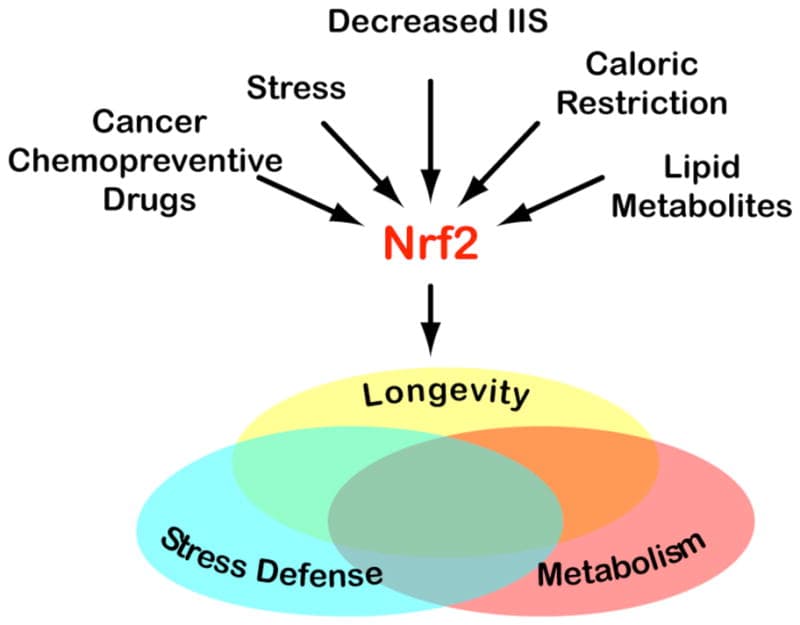
Sulforaphane: An isothiocyanate from cruciferous vegetables (broccoli sprouts are the highest source) with established HDAC inhibitory activity. Sulforaphane activates nuclear factor erythroid 2-related factor 2 (Nrf2), which upregulates antioxidant defenses, and independently inhibits HDAC activity, increasing histone acetylation at gene promoters. Sulforaphane's HDAC inhibitory potency is lower than pharmaceutical inhibitors but it crosses the blood-brain barrier more readily than sodium butyrate and has been detected in brain tissue after oral administration. R
Resveratrol: A polyphenol from grapes and red wine that has complex effects on histone modification. Resveratrol activates Sirtuin 1 (SIRT1), a Class III HDAC (NAD+-dependent deacetylase), which has different substrate specificity than the Class I HDACs most relevant to extinction. At the same time, resveratrol inhibits some Class I HDAC activity. Its net effect on fear extinction is not well characterized, but it is included here because it is frequently discussed in the context of epigenetic modulation. R
Exercise: Voluntary physical exercise increases histone H3 acetylation in the hippocampus and prefrontal cortex in rodents, in a pattern consistent with enhanced neuroplasticity. R Exercise also increases hippocampal BDNF expression, which is the downstream effector of extinction-induced histone acetylation. Several clinical studies show that aerobic exercise before or after exposure therapy sessions improves PTSD outcomes, consistent with an exercise-mediated epigenetic priming mechanism.
Sleep: Sleep deprivation impairs extinction consolidation. Memory consolidation, including extinction memory consolidation, is substantially dependent on slow-wave and REM sleep during which hippocampal and prefrontal plasticity genes are re-expressed and synaptic changes are stabilized. The practical implication is that post-extinction sleep is not optional if lasting fear inhibition is the goal. R
Where PTSD Treatment Currently Stands
PTSD affects approximately 7 to 8% of the general population and up to 30% of combat veterans. First-line pharmacological treatments are serotonin reuptake inhibitors (SSRIs), which have only a 50 to 60% response rate and primarily manage symptoms rather than addressing the underlying fear memory mechanisms. R
Exposure-based therapies (prolonged exposure, EMDR (eye movement desensitization and reprocessing), cognitive processing therapy) are more effective for many patients but are limited by:
- Context-dependence of extinction (fear returns in new environments)
- Spontaneous recovery over time
- Treatment resistance in patients with remote or severe trauma
- High dropout rates due to the distress of exposure
The HDAC inhibitor approach represents a pharmacological augmentation strategy: administered in conjunction with exposure therapy during the extinction or reconsolidation window, not as a standalone treatment. The working model is that the HDAC inhibitor epigenetically primes the brain to consolidate the extinction session more deeply, converting a weak or temporary fear reduction into a durable fear inhibitory memory.
This approach has not yet reached human clinical trials specifically for fear extinction in PTSD. The obstacles include:
- Current HDAC inhibitors approved for human use (vorinostat, romidepsin, belinostat, panobinostat) are approved for oncology and carry significant side effect profiles at the doses used for cancer
- Developing HDAC inhibitors with CNS penetration, Class I selectivity, and tolerable side effect profiles at lower doses is an active area
- Timing and dosing windows for maximal extinction benefit in humans are not yet established
The most immediately translatable approach in clinical practice is pairing exposure therapy with the natural HDAC modulators described above (butyrate-raising dietary strategies, sulforaphane, aerobic exercise, optimized sleep) to support the epigenetic environment in which extinction consolidation occurs. R
Mechanisms Of Action
Simple:
- Fear extinction is new learning, not memory erasure. The original fear memory persists and can return; it must be actively suppressed by a competing extinction memory.
- The brain circuit for extinction runs from the infralimbic cortex (IL) through intercalated cells (ITCs) to the central amygdala (CeA): IL activation silences CeA fear output by activating inhibitory ITC neurons.
- Extinction memory consolidation requires histone acetylation in the hippocampus and infralimbic cortex, specifically histone H4 acetylation at the BDNF exon IV promoter, to drive the BDNF expression needed for synaptic plasticity.
- HDAC2 is the key epigenetic regulator. In recent memories, retrieval triggers S-nitrosylation of HDAC2 that releases it from chromatin, opening a neuroplasticity window during which extinction training is most effective.
- In remote (older) memories, this HDAC2 S-nitrosylation does not occur, the plasticity window stays closed, and extinction training fails to consolidate properly. This is why PTSD is harder to treat the longer it has been present.
- HDAC inhibitors, when given after extinction training or during the reconsolidation window, increase histone acetylation and extend or restore the neuroplasticity window, converting weak extinction experiences into durable long-term fear inhibition.
- HDAC1 and HDAC2, not HDAC3, are the relevant isoforms for fear extinction consolidation.
- Natural HDAC inhibitors (butyrate from dietary fiber, sulforaphane from broccoli sprouts) and lifestyle factors (aerobic exercise, optimized sleep) support the epigenetic environment for extinction without pharmaceutical intervention.
Advanced:
- HDAC2 S-nitrosylation cascade: Memory retrieval activates NMDA receptors at hippocampal CA1 synapses, triggering calcium influx that activates neuronal nitric oxide synthase (nNOS). nNOS-derived nitric oxide reacts with cysteine residues 262 and 274 of the HDAC2 protein, causing S-nitrosylation. This post-translational modification induces a conformational change that releases HDAC2 from chromatin-associated co-repressor complexes (NuRD complex, Sin3A), removing the enzymatic barrier to histone acetylation at immediate early gene (IEG) and neuroplasticity gene promoters. The resulting H3K9/14 acetylation at the c-Fos promoter drives c-Fos transcription, which initiates a gene expression cascade including BDNF, Arc (activity-regulated cytoskeleton-associated protein), and other plasticity genes that enable dendritic spine remodeling, AMPA receptor insertion, and new synaptic connections needed to store the extinction trace. R
- Differential BDNF promoter regulation: The BDNF gene has at least nine distinct 5' non-coding exons, each with its own promoter, converging on a common coding exon. Fear conditioning preferentially upregulates H3 acetylation at BDNF promoters I and IV in the lateral amygdala, increasing BDNF exon IV transcripts that support fear memory consolidation. Fear extinction upregulates H4 acetylation at BDNF promoter IV specifically in the infralimbic cortex and hippocampus. These distinct histone marks (H3 vs H4) recruit different chromatin remodeling complexes and transcription factors, suggesting that fear and extinction utilize biochemically distinct BDNF induction pathways in distinct neural circuits. VPA and NMDA receptor activation predominantly increase exon IV-specific BDNF transcripts, mechanistically linking HDAC inhibition and glutamate receptor signaling to the same extinction-relevant BDNF pathway. R
- Reconsolidation window boundary conditions: The reconsolidation window is triggered by brief CS-only presentation (typically 1 to 2 CS exposures) and lasts approximately 6 hours, during which the memory is labile and protein synthesis-dependent. Boundary conditions that determine whether retrieval triggers reconsolidation versus extinction include: number of CS presentations (few = reconsolidation, many = extinction), time since original conditioning, memory strength, and the presence of new information during retrieval. During the reconsolidation window, AMPA receptor-mediated currents increase transiently relative to NMDA-mediated currents, reflecting a state of heightened synaptic flexibility. HDAC inhibitors applied during this window maintain H3K9/14 acetylation at neuroplasticity gene promoters for longer than retrieval alone achieves, effectively extending the lability window and giving extinction training more time to incorporate into the updating memory trace. R
- Engram competition model: Fear and extinction are encoded in partially overlapping but distinct neuronal ensembles (engrams) in the BLA and mPFC. Fear engram neurons in the BLA are activated by the CS and project to CeA to drive fear output. Extinction engram neurons in the BLA receive inputs from IL and project to ITC, driving inhibition of fear output. Long-term fear inhibition depends on IL-to-BLA connectivity being strong enough to activate extinction engram neurons in preference to fear engram neurons when the CS is encountered. HDAC inhibition increases BDNF expression in the IL-hippocampal network, which enhances axonal sprouting and dendritic spine density in the IL-to-BLA pathway, structurally strengthening the extinction engram's competitive advantage over the fear engram. R
Genetics
BDNF Val66Met (rs6265): The most studied genetic variant in fear extinction. The Val66Met polymorphism (valine to methionine substitution at codon 66) impairs activity-dependent secretion of BDNF from neurons. Humans and mice carrying the Met allele show impaired fear extinction: they acquire fear normally but fail to consolidate extinction memory adequately, producing higher rates of return of fear after exposure therapy. R Because HDAC inhibition enhances extinction specifically through BDNF upregulation, the BDNF Val66Met variant may predict who benefits most from HDAC inhibitor augmentation of exposure therapy. Met allele carriers represent a subgroup in whom standard exposure therapy is most likely to fail and in whom epigenetic augmentation strategies may be most needed.
FKBP5 (rs1360780, rs9296158, rs3800373): FK506-binding protein 5 (FKBP5) is a co-chaperone of the glucocorticoid receptor that regulates stress hormone sensitivity. FKBP5 risk variants increase glucocorticoid receptor resistance, producing a state of higher chronic cortisol exposure. Elevated glucocorticoids impair extinction by enhancing BLA activity and suppressing IL function, and by promoting HDAC recruitment to neuroplasticity gene promoters in the hippocampus, reducing basal histone acetylation in extinction-relevant circuits. FKBP5 risk variants are strongly associated with PTSD risk after trauma exposure, consistent with the mechanistic prediction that impaired extinction is a key intermediate phenotype. R The interaction between FKBP5 variants and childhood trauma is one of the most replicated gene-environment interactions in psychiatric genetics.
NR3C1 (glucocorticoid receptor gene): Methylation of NR3C1 promoter regions, particularly the 1F promoter, is increased by early life adversity and reduces glucocorticoid receptor expression. Reduced glucocorticoid receptor expression alters HPA axis feedback, producing chronic stress dysregulation that impairs extinction via the mechanisms described under FKBP5. NR3C1 methylation is detectable in peripheral blood and has been proposed as a biomarker for PTSD risk and treatment response, though peripheral methylation may not fully reflect central nervous system methylation patterns. R
More Research
- The reconsolidation-extinction window in humans has been replicated and validated, but the optimal number of CS presentations to trigger reconsolidation without triggering extinction varies across individuals and is currently estimated clinically rather than measured. Developing objective biomarkers for the reconsolidation window (such as pupil dilation response, skin conductance patterns, or EEG signatures) is an active area of research that could allow clinicians to time HDAC inhibitor administration precisely. R
- MDMA (3,4-methylenedioxymethamphetamine)-assisted psychotherapy for PTSD, which is in Phase 3 clinical trials, may work in part through epigenetic mechanisms including temporary reduction in fear-related amygdala reactivity that mirrors a pharmacologically induced reconsolidation window, combined with enhanced extinction consolidation via BDNF upregulation. The mechanistic overlap with HDAC inhibitor-based approaches is not coincidental and suggests both interventions may ultimately target the same synaptic plasticity pathways through different upstream mechanisms. R
- Propranolol (a beta-adrenergic receptor antagonist) is the most studied pharmacological reconsolidation-interfering agent in humans. Administered before CS retrieval, propranolol reduces reconsolidation of the fear memory by blocking norepinephrine-mediated memory strengthening during the lability window. Combining propranolol (to weaken the fear memory during reconsolidation) with an HDAC inhibitor (to strengthen the extinction memory after the same session) is a theoretically attractive dual intervention that has not yet been tested in humans. R
- The gut-brain axis connection to fear extinction is emerging: butyrate-producing gut microbiota have been shown to influence fear extinction behavior in rodents, with germ-free mice (lacking gut microbiota and therefore gut-derived butyrate) showing impaired extinction consolidation. Reconstituting gut microbiota or supplementing with sodium butyrate partially rescues extinction deficits in germ-free animals, suggesting that the gut microbiome actively maintains the epigenetic environment of the brain's fear extinction circuitry through circulating SCFA signaling. R
- The distinction between HDAC inhibition and HAT (histone acetyltransferase) activation is clinically important. Both increase histone acetylation, but via opposite routes. HAT inhibition during extinction consolidation impairs extinction retention (consistent with a requirement for acetyltransferase activity to write the extinction mark). HDAC inhibition enhances extinction retention. Current drug development focuses on HDAC inhibitors rather than HAT activators because the HDAC enzyme active sites are more druggable, but both targets are mechanistically valid. R
Jacob Gordon
INHC, FMT-C
Board Certified Health Coach
I spent years battling unexplained chronic illness before discovering biohacking, epigenetics, and functional medicine. Now I share that research at MyBioHack to help others find their own answers.
Book a ConsultationRelated Protocols & Supplements
Deep-dive chapters and recommended supplements for this topic
Lion's Mane
1000mg/day
Omega-3 (DHA)
2g/day
Phosphatidylserine
100mg 3x/day