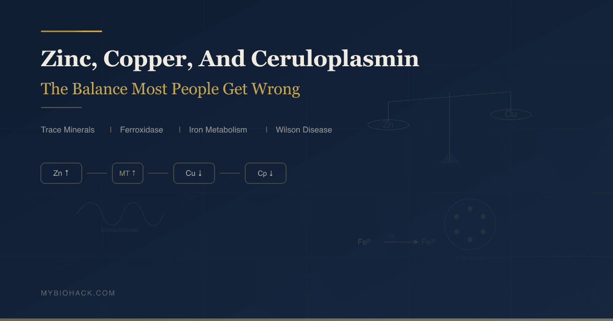
Zinc, Copper, And Ceruloplasmin: The Balance Most People Are Getting Wrong
By Jacob Gordon, INHC, FMT-CThis article contains affiliate links. As an Amazon Associate, MyBioHack earns from qualifying purchases at no extra cost to you. We only link products we research and stand behind.
Zinc and copper are the most interdependent minerals in human biochemistry, and the relationship between them is one of the most clinically underrecognized and most commonly disrupted by supplementation.
In this post, we will discuss what zinc and copper each do, how ceruloplasmin connects them to iron metabolism, what goes wrong when the balance is disrupted in either direction, how long-term zinc supplementation silently depletes copper, what Wilson disease and aceruloplasminemia reveal about the system, and how to test and correct the balance.
Basics Of Zinc, Copper, And Their Relationship
Zinc and copper are both essential trace minerals and both cofactors for critical enzymes throughout the body.
They are absorbed in overlapping pathways in the proximal small intestine, and they compete for the same intracellular binding protein: metallothionein.
This competition is not incidental. It is a fundamental feature of how the body regulates both minerals simultaneously.
When zinc is elevated, copper is suppressed. When copper is elevated, zinc is affected. The ratio between them matters as much as the absolute level of either one. R
The normal serum zinc:copper ratio is approximately 0.7-1.0 (zinc:copper in mg/L) in most reference ranges.
Disruption of this ratio is associated with immune dysfunction, anemia, neurological symptoms, and altered iron metabolism, depending on which direction the balance tips.
Most people supplement zinc without knowing they are simultaneously suppressing copper.
This is not a theoretical concern. It is documented in the clinical literature through cases of sideroblastic anemia, neutropenia, and myelopathy directly caused by zinc-induced copper deficiency from over-the-counter zinc supplements and zinc-containing denture adhesives. R
What Zinc Does
Zinc is the second most abundant trace element in the human body after iron, present at approximately 2-3 grams total. R
It is a cofactor or structural component in over 300 enzymes, making it involved in more biochemical reactions than any other trace mineral. R
The major functions of zinc include: (not exclusive list)
- Alcohol dehydrogenase (alcohol metabolism and detoxification)
- Carbonic anhydrase (acid-base balance, CO2/bicarbonate interconversion, essential for respiration)
- Cu/Zn Superoxide Dismutase (SOD1) (the primary cytosolic antioxidant enzyme, converts superoxide radicals to hydrogen peroxide and oxygen; discussed further in the Copper section)
- DNA polymerases (DNA replication, repair, and recombination; zinc stabilizes polymerase structure and enhances activity)
- Immune cell development and signaling (T-cell maturation, NK cell activity, neutrophil function; zinc deficiency impairs virtually every arm of both innate and adaptive immunity)
- Insulin production and storage (insulin is stored as a hexamer with zinc in pancreatic beta cells; zinc is needed for correct insulin crystallization and secretion)
- Matrix metalloproteinases (MMPs) (wound healing, tissue remodeling, collagen degradation)
- Testosterone synthesis (zinc inhibits aromatase, preventing testosterone conversion to estrogen; zinc deficiency increases aromatase activity and reduces testosterone; LH and FSH secretion from the pituitary are impaired by zinc deficiency) R
- Thymulin (a thymic hormone that is zinc-dependent; thymulin is inactive without zinc and is required for T-cell maturation and function)
Common symptoms of zinc deficiency include: (not exclusive list)
- Alopecia (diffuse hair loss)
- Delayed wound healing
- Diminished sense of taste and smell (hypogeusia, hyposmia)
- Frequent infections
- Hypogonadism and low testosterone
- Impaired growth in children
- Night blindness (zinc is required for retinol binding protein synthesis and vitamin A transport)
- Poor appetite
- Skin changes (acrodermatitis enteropathica in severe genetic deficiency; eczema-like changes in acquired deficiency)
Natural sources of zinc by concentration: (not exclusive list)
- Beef (5.5mg/100g)
- Cashews (5.6mg/100g)
- Crab (6.5mg/100g)
- Lamb (6.7mg/100g)
- Oysters (highest natural source, 74mg/100g)
- Pepitas/pumpkin seeds (7.6mg/100g)
Phytates in grains and legumes bind zinc and substantially reduce its bioavailability. R
This is why vegetarians and vegans have lower circulating zinc despite adequate dietary intake in grams.
What Copper Does
Copper is an essential cofactor for enzymes spanning antioxidant defense, energy production, connective tissue synthesis, iron metabolism, and neurotransmitter function. R
The major copper-dependent enzymes include: (not exclusive list)
- Ceruloplasmin (ferroxidase; carries over 95% of plasma copper; essential for iron metabolism, discussed in depth below) R
- Cu/Zn Superoxide Dismutase (SOD1) (requires both copper AND zinc for activity; copper is the catalytic active site; zinc stabilizes protein structure; the dismutation reaction itself is copper-mediated) R
- Cytochrome c oxidase (Complex IV) (the terminal enzyme of the mitochondrial electron transport chain; copper is essential for the final electron transfer to oxygen in oxidative phosphorylation; copper deficiency impairs mitochondrial energy production)
- Dopamine beta-hydroxylase (DBH) (converts dopamine to norepinephrine; copper deficiency directly impairs norepinephrine synthesis and adrenergic neurotransmission) R
- Lysyl oxidase (LOX) (cross-links collagen and elastin, providing structural integrity to connective tissue, blood vessels, and bone; copper deficiency impairs wound healing, aortic integrity, and bone strength)
- Peptidylglycine alpha-amidating monooxygenase (PAM) (required for activation of several neuropeptides including substance P, VIP, oxytocin, and CRH)
- Tyrosinase (melanin synthesis in melanocytes; copper deficiency causes depigmentation of skin and hair) R
Common symptoms of copper deficiency include: (not exclusive list)
- Anemia unresponsive to iron therapy (hypochromic, microcytic or normocytic)
- Ataxic gait and sensory ataxia (copper deficiency myelopathy)
- Depigmentation of hair and skin (low melanin from tyrosinase impairment)
- Fatigue and weakness
- Frequent infections (neutropenia)
- Joint pain
- Neurological symptoms (peripheral neuropathy, myelopathy mimicking B12 deficiency)
- Neutropenia (low absolute neutrophil count)
- Osteoporosis (impaired lysyl oxidase, reduced collagen crosslinking)
Natural sources of copper by concentration: (not exclusive list)
- Beef liver (highest practical source, 14mg/100g)
- Black pepper (dried, 1.3mg/100g)
- Cashews (2.2mg/100g)
- Dark chocolate (1.8mg/100g)
- Lentils (0.75mg/100g)
- Oysters (4.5mg/100g)
- Sesame seeds (4.1mg/100g)
- Shiitake mushrooms (0.9mg/100g)
Liver is the single most copper-dense commonly available food.
The RDA for copper is 900mcg/day for adults.
Ceruloplasmin: The Ferroxidase At The Center
Ceruloplasmin (Cp) is a copper-containing glycoprotein synthesized primarily by hepatocytes and secreted into blood, where it carries over 95% of all copper in plasma. R
Normal serum ceruloplasmin is 20-40 mg/dL.
Ceruloplasmin contains six copper atoms per molecule and belongs to the multicopper oxidase family: proteins that use copper to couple substrate oxidation with the four-electron reduction of oxygen to water.
Despite containing the vast majority of plasma copper, ceruloplasmin does not play the primary role in copper transport or distribution to tissues.
Its central physiological role is as a ferroxidase: it oxidizes ferrous iron (Fe2+, the toxic reduced form) to ferric iron (Fe3+, the non-toxic oxidized form). R
This matters enormously because:
Only Fe3+ can bind to transferrin, the blood protein that transports iron to tissues. R
Without ceruloplasmin's ferroxidase activity, ferrous iron cannot be loaded onto transferrin for systemic delivery.
Iron then cannot exit cells efficiently via ferroportin (the iron efflux transporter), leading to intracellular iron accumulation.
The result is simultaneous intracellular iron overload AND functional iron deficiency in circulation, creating the paradoxical picture of high ferritin with anemia and low transferrin saturation that is characteristic of ceruloplasmin deficiency. R
Ceruloplasmin in the brain is different from ceruloplasmin in blood.
In the CNS, astrocytes express a GPI-anchored form of ceruloplasmin (GPI-Cp) on their cell surface.
This membrane-bound form is the primary ferroxidase in the brain, and it is absolutely essential for iron homeostasis in the CNS. R
GPI-Cp on astrocytes oxidizes ferrous iron exported from neurons via ferroportin, loads it onto transferrin, and recycles it back to neurons via the transferrin receptor.
Without functional GPI-Cp, ferrous iron accumulates in astrocytes, causing lipid peroxidation and oxidative damage to the very cells that are supposed to support and protect neurons. R
This mechanism explains why aceruloplasminemia causes neurodegeneration despite being a disorder of copper and ceruloplasmin: the failure is ultimately an iron accumulation problem in the brain. R
Ceruloplasmin is also a positive acute-phase reactant, meaning it rises with inflammation, infection, pregnancy, estrogen use, and malignancy.
This is clinically important: inflammation can normalize a low ceruloplasmin, masking underlying copper deficiency and creating a false-negative result. R
Always interpret ceruloplasmin in context of inflammatory markers and serum copper together.
How Zinc Depletes Copper: The Metallothionein Mechanism
This is the mechanism behind dozens of documented clinical cases of copper deficiency from zinc supplementation, and it is almost certainly happening at a subclinical level in a much larger population of supplement users.
Metallothionein (MT) is a small cysteine-rich intracellular protein in enterocytes (intestinal absorptive cells) that binds trace metals. R
When zinc intake is elevated, enterocytes increase metallothionein synthesis as a homeostatic response, because MT sequesters excess zinc and prevents its absorption. R
The problem: metallothionein has a much higher binding affinity for copper than for zinc.
When dietary copper arrives in the gut in the presence of elevated metallothionein, the copper binds to metallothionein before zinc does.
Copper-bound metallothionein is trapped inside the enterocyte.
As the enterocyte naturally undergoes cell turnover and is shed into the intestinal lumen (normal process, every 3-5 days), the copper bound to metallothionein is lost in the feces rather than being absorbed into the bloodstream. R
Systemic copper absorption falls. Over weeks to months, serum copper and ceruloplasmin decline.
This mechanism explains several critically important clinical observations:
- The clinical manifestations of zinc excess are actually the manifestations of copper deficiency: anemia, neutropenia, and myelopathy.
- Even normal-appearing serum zinc levels can indicate ongoing copper depletion, because the excess zinc that drove up metallothionein may have already been excreted but the metallothionein production is still suppressing copper absorption.
- In documented cases, stopping zinc supplements and starting oral copper fails to correct the copper deficiency until excess zinc is eliminated from the body, because elevated metallothionein continues to block copper absorption as long as zinc excess persists. R
- In some cases, intravenous copper was required to bypass the blocked gut absorption entirely. R
How much zinc is too much?
Intake above 40mg/day regularly is considered the threshold for concern. R
Many common OTC supplements contain 25-50mg of zinc per dose.
Long-term use at these doses without copper co-supplementation is a real clinical risk.
Zinc-containing denture adhesives have caused copper deficiency and myelopathy from dermal absorption of zinc, in patients who used them heavily for years. R
The ratio to aim for in supplementation: Most practitioners use a zinc:copper ratio of approximately 8-15:1 (by dose in mg) when supplementing both.
If supplementing 15mg of zinc daily, approximately 1-2mg of copper daily is appropriate to maintain balance.
Conditions Linked To Zinc-Copper Imbalance
Zinc Excess / Copper Deficiency
Copper deficiency myelopathy is the most severe neurological consequence of zinc-induced copper depletion.
It presents as a progressive myelopathy (spinal cord disease) with spastic gait, sensory ataxia, and absent or diminished deep tendon reflexes with Babinski signs, closely mimicking subacute combined degeneration from vitamin B12 deficiency. R
Vitamin B12 and copper deficiency can coexist and should always be tested together when either is suspected. R
Sideroblastic anemia and neutropenia are the most common hematological presentations.
The anemia is iron-unresponsive (a critical diagnostic clue), because copper deficiency impairs iron absorption from the gut and iron release from the reticuloendothelial system.
Bone marrow biopsy shows ringed sideroblasts and vacuolization of erythroid and myeloid precursors, which can be misdiagnosed as myelodysplastic syndrome (MDS). R
Unlike MDS, copper deficiency-related marrow changes are fully reversible with copper restoration.
Low testosterone can occur with long-term zinc-induced copper depletion if it impairs cytochrome c oxidase and dopamine-beta-hydroxylase function, affecting adrenergic signaling pathways relevant to testosterone synthesis. Paradoxically, zinc deficiency also causes low testosterone through a different mechanism (low aromatase inhibition).
Dysbiosis is both a cause and consequence of trace mineral imbalance. Gut pathogens and SIBO alter zinc and copper absorption independently of supplementation.
Copper Excess / Aceruloplasminemia
Aceruloplasminemia is a rare autosomal recessive disorder caused by loss-of-function mutations in the ceruloplasmin gene, resulting in complete absence of ceruloplasmin ferroxidase activity. R
Despite being a copper-containing protein disorder, the clinical syndrome is primarily one of iron accumulation, not copper toxicity.
Massive iron deposition occurs in the brain (basal ganglia, dentate nuclei, thalamus, cortex), liver, pancreas, and retina. R
Onset is typically in the 4th-5th decade with:
- Blepharospasm, orolingual dystonia, chorea, dysarthria, ataxia
- Parkinsonism
- Cognitive decline and dementia
- Diabetes mellitus (pancreatic iron deposition damages beta cells)
- Retinal pigmentary degeneration
- Microcytic anemia with paradoxically high ferritin and low transferrin saturation
On MRI, aceruloplasminemia shows the most extreme T2 hypointensity (iron signal) in the basal ganglia of any condition, surpassing Wilson disease. R
Wilson Disease is the mirror image disorder: an autosomal recessive defect in ATP7B, the copper transporter in hepatocyte trans-Golgi network.
ATP7B normally incorporates copper into apoceruloplasmin to form holoceruloplasmin, and mediates biliary excretion of excess copper. R
In Wilson disease, ATP7B dysfunction means copper cannot be incorporated into ceruloplasmin and cannot be excreted into bile.
Free (non-ceruloplasmin-bound) copper accumulates and is toxic.
Serum ceruloplasmin is typically low in Wilson disease because non-coppered apoceruloplasmin is unstable and rapidly degraded. R
Paradoxically, treatment of Wilson disease includes zinc supplementation to induce metallothionein in the gut and block copper absorption, the exact same mechanism that causes copper deficiency in otherwise healthy supplement users. R
Neurodegeneration and Alzheimer's/Parkinson's connection: Ceruloplasmin ferroxidase activity is reduced in the CSF of Parkinson's disease patients. Ceruloplasmin levels are reduced in the cortex in Alzheimer's disease. R
Both diseases are associated with iron accumulation in the substantia nigra (Parkinson's) and amyloid plaques (Alzheimer's).
Impaired ceruloplasmin ferroxidase activity may contribute to neurodegenerative iron accumulation in both conditions.
Mast cell activation is influenced by copper status. Histamine N-methyltransferase (HNMT), which degrades histamine, is a methylation enzyme affected by copper status. Copper-dependent enzymes are also involved in catecholamine synthesis that modulates mast cell reactivity.
How To Balance Zinc And Copper
1. Assess Before Supplementing
Test serum copper, serum zinc, and serum ceruloplasmin together before beginning any supplementation protocol.
Serum copper and ceruloplasmin must be interpreted together: ceruloplasmin carries most plasma copper, so low ceruloplasmin almost always means low functional copper (with the caveat that inflammation can normalize ceruloplasmin even when copper is depleted).
If ceruloplasmin is low but the inflammatory picture is unclear, requesting a serum copper alongside it clarifies whether the low ceruloplasmin reflects genuine copper deficiency or is being masked by inflammation.
2. Correct Copper Deficiency First
If copper is low and zinc is elevated, the priority is reducing zinc intake and restoring copper.
Copper Glycinate or Copper Bisglycinate at 1-3mg/day is appropriate for mild to moderate deficiency in a patient who can absorb oral supplements.
If GI absorption is compromised (malabsorption, bariatric surgery, or elevated metallothionein blocking absorption), IV copper may be required to bypass the gut. R
Do not supplement copper aggressively in isolation without testing, as copper excess (Wilson disease phenotype without the genetic defect) is possible and damaging.
3. Supplement Zinc With A Co-Administered Ratio
When zinc supplementation is indicated (infection, wound healing, testosterone support, immune function, taste disturbances), always include copper at approximately 1mg of copper per 10-15mg of zinc.
Zinc Glycinate or Zinc Bisglycinate are the best-absorbed forms with the least GI side effects.
Zinc + Copper Combined Supplement products are available and simplify compliance.
Zinc picolinate and zinc citrate are also well-absorbed.
Zinc oxide (common in cheaper supplements) has significantly lower bioavailability.
4. Support Ceruloplasmin Production
Ceruloplasmin is synthesized in the liver and requires both adequate copper status and functional hepatic biosynthesis.
Liver health is a prerequisite for normal ceruloplasmin levels. Milk thistle (silymarin) and TUDCA support hepatic function relevant to ceruloplasmin synthesis.
Ceruloplasmin synthesis also requires adequate vitamin A (retinol form), as retinol modulates copper metabolism in the liver.
Vitamin C enhances ferroxidase function and may support ceruloplasmin activity indirectly.
5. Dietary Copper From Food
Prioritizing copper-rich foods is the most sustainable approach for most people.
Beef liver 1-2 times per week provides substantial copper in a highly bioavailable form.
Oysters provide both zinc and copper at meaningful amounts.
Dark chocolate (high cocoa) and cashews are practical everyday copper sources.
6. Address Absorption Factors
Dysbiosis reduces copper absorption through multiple pathways including altered gut pH, impaired enterocyte function, and competitive binding by gut organisms.
Malabsorptive GI surgery (Roux-en-Y gastric bypass has a 9-10% prevalence of post-operative copper deficiency) is a major risk factor that requires ongoing monitoring. R
Celiac disease and other causes of villous atrophy significantly reduce copper absorption.
What To Stay Away From
- High-dose zinc without copper co-administration (anything above 25mg/day of zinc without 1-2mg of copper coadministered is creating a depletion risk over time; the clinical literature documents copper deficiency from this pattern)
- Zinc-containing denture adhesives used chronically (dermal zinc absorption from heavy denture adhesive use has caused copper deficiency myelopathy; report this to dental patients explicitly) R
- Interpreting ceruloplasmin in isolation (ceruloplasmin is an acute-phase reactant; it rises with inflammation, oral contraceptive use, pregnancy, and valproate use, potentially masking true copper deficiency; always pair it with serum copper and inflammatory markers like CRP or ferritin) R
- Iron supplementation without ruling out ceruloplasmin deficiency (iron therapy will not correct the anemia of copper deficiency; treating with iron when the underlying issue is copper deficiency delays correct diagnosis and exposes the patient to excess iron without benefit)
- Copper supplementation in Wilson disease (copper accumulation is the disease; supplementing copper in an undiagnosed Wilson disease patient is directly harmful)
- Long-term high-dose zinc during active infection or post-COVID recovery (zinc supplementation surged post-COVID and many patients continued high doses indefinitely without copper monitoring; this is a copper deficiency incubation period)
- Mega-dose zinc lozenges for extended cold management (total daily zinc from lozenges can easily exceed 100mg/day; this is effective short-term for cold duration but should not be continued for more than 5-7 days without copper co-supplementation)
- Neglecting copper status in vegetarians and vegans (plant-based diets are lower in bioavailable copper than meat-based diets; compound this with common zinc supplementation in this demographic and the depletion risk is significant)
Testing
First-Line Testing
Serum Zinc and Serum Copper should always be ordered together when evaluating either mineral. Individual results without the paired measurement are clinically incomplete.
Serum Ceruloplasmin should be ordered simultaneously.
Reference ranges (approximate):
- Serum copper: 0.75-1.45 mcg/mL (75-145 mcg/dL)
- Serum ceruloplasmin: 20-40 mg/dL
- Serum zinc: 0.66-1.0 mcg/mL
Copper less than 7.9 mcmol/L is considered severe deficiency warranting specialist referral. R
The Nutrient Zoomer from Vibrant Wellness includes serum copper, zinc, and ceruloplasmin as part of its comprehensive micronutrient panel, alongside the vitamins and minerals that context these results.
Individual markers are also available separately: Copper and Zinc via Fullscript Quest Diagnostics.
Second-Line Testing
CBC with Differential: look for anemia and neutropenia as the hematological signature of copper deficiency.
Anemia that is unresponsive to iron therapy and is accompanied by neutropenia should prompt immediate copper evaluation.
24-hour urine copper excretion is useful when Wilson disease (copper excess) or non-ceruloplasmin-bound copper accumulation is suspected. Elevated in active Wilson disease, reduced in copper deficiency.
Iron studies Foundation Zoomer: ferritin, transferrin saturation, serum iron, and TIBC to assess functional iron status alongside the copper-ceruloplasmin picture.
The paradox of high ferritin with low transferrin saturation and anemia (the aceruloplasminemia pattern) should trigger ceruloplasmin testing.
Neurological Evaluation
When copper deficiency myelopathy is suspected, MRI of the cervical and thoracic spinal cord shows T2 hyperintensity in the dorsal columns, identical in appearance to subacute combined degeneration from B12 deficiency. R
The Neural Zoomer from Vibrant Wellness assesses autoantibodies and markers of neuroinflammation relevant to patients with neurological symptoms from trace mineral dysregulation.
Mechanisms Of Action
Simple:
- Zinc and copper compete for the same binding protein (metallothionein) in gut cells; when zinc is high, metallothionein preferentially grabs copper and carries it out of the body when the gut cell is shed.
- Copper is required to build ceruloplasmin, the protein that converts toxic ferrous iron (Fe2+) into the safe ferric form (Fe3+) that can ride on transferrin and get where it needs to go.
- Without ceruloplasmin, iron cannot leave cells via the ferroportin transporter, so iron builds up inside cells even while blood levels suggest iron deficiency.
- In the brain, astrocytes use a surface form of ceruloplasmin to recycle iron between themselves and neurons; when this breaks down, iron accumulates in astrocytes, causing oxidative damage that destroys neuronal support.
- Cu/Zn-SOD1 requires both copper AND zinc to function; the copper center does the dismutation chemistry; the zinc center holds the protein in the right shape. Lose either and your primary cytosolic antioxidant enzyme fails.
Advanced:
- Metallothionein induction cascade: Zinc enters the enterocyte via ZIP4 (SLC39A4), elevating intracellular Zn2+. This activates MTF-1 (metal-responsive transcription factor-1), which binds metal response elements (MREs) in the metallothionein promoter and drives MT1 and MT2 expression. The resulting MT proteins have 20 cysteine residues that bind up to 7 zinc atoms or a mixture of metals. MT's binding affinity for Cu is approximately 100-fold greater than for Zn (MT Kd for Cu ~10^-18M vs Kd for Zn ~10^-12M). Dietary copper entering the enterocyte is immediately sequestered by Cu-preferring MT binding sites, preventing transfer to the basolateral copper transporter CTR1 that would export copper to portal circulation. R
- Ceruloplasmin copper incorporation: Holoceruloplasmin is synthesized exclusively in hepatocytes. In the trans-Golgi network, ATP7B transports copper from the cytoplasm to the lumenal side of the Golgi, where the copper chaperone ATOX1 delivers it to ATP7B, which then loads it into nascent apoceruloplasmin via sequential metalation of its six copper-binding sites. Without ATP7B function (Wilson disease), apoceruloplasmin cannot be metalated and is rapidly degraded, explaining the low ceruloplasmin in Wilson disease despite excess hepatic copper. R
- Iron efflux and ferroxidase coupling: Iron stored inside macrophages, hepatocytes, and enterocytes exits via ferroportin (FPN1/SLC40A1), which specifically transports ferrous iron (Fe2+). At the cell surface, membrane-bound ceruloplasmin (GPI-Cp in astrocytes, soluble Cp in hepatocytes) immediately oxidizes Fe2+ to Fe3+. Only Fe3+ can bind to transferrin (TF) via its iron-binding lobes. This oxidation step is the rate-limiting bottleneck for iron mobilization and systemic delivery. Without ceruloplasmin activity, ferrous iron exits ferroportin but cannot be loaded onto transferrin, stalling in the interstitium. Hepcidin then upregulates in response to the apparent iron excess, induces ferroportin internalization and degradation, and traps even more iron intracellularly. This explains the low-transferrin-saturation, high-ferritin presentation of aceruloplasminemia. R
- Cu/Zn-SOD1 (SOD1) mechanism: SOD1 is a 32kDa homodimer with one copper and one zinc per subunit. The copper ion cycles between Cu2+ and Cu+ during catalysis: Cu2+-SOD + O2- → Cu+-SOD + O2 (copper reduced), then Cu+-SOD + O2- + 2H+ → Cu2+-SOD + H2O2 (copper re-oxidized, superoxide reduced to H2O2). Zinc does not participate in this redox cycle; it stabilizes the protein's Greek key beta-barrel structure and maintains the shape of the active site for copper positioning. CCS (copper chaperone for SOD1) specifically delivers copper to nascent SOD1 and also forms the disulfide bond that locks the active conformation. Loss of either copper or zinc impairs SOD1 function, reducing cytosolic superoxide dismutation. R
- Aceruloplasminemia iron-astrocyte-neuron cycle disruption: In normal brain, astrocytes export Fe3+ bound to transferrin across the interstitium to neurons, where TfR1 (transferrin receptor 1) internalizes the Fe3+-transferrin complex. GPI-Cp on the astrocyte surface is required to oxidize intracellular Fe2+ (exported from astrocytes via ferroportin) to Fe3+ for loading onto transferrin. In aceruloplasminemia, Fe2+ exits astrocytes via ferroportin but cannot be oxidized, so it fails to bind transferrin and instead accumulates in the astrocyte interstitium and cytoplasm. The non-transferrin-bound iron (NTBI) undergoes Fenton chemistry (Fe2+ + H2O2 → Fe3+ + OH• + OH-), generating the most potent reactive oxygen species in biology. Astrocytes, overwhelmed by oxidative damage, lose their capacity to support neurons. Neurons, starved of transferrin-bound iron, eventually begin taking up NTBI via alternative pathways that further increase their oxidative burden. R
Genetics
ATP7B (Wilson Disease)
ATP7B encodes the ATPase copper transporter ATP7B on chromosome 13q14.3.
ATP7B is expressed primarily in hepatocytes and functions in the trans-Golgi network to incorporate copper into ceruloplasmin and to traffic excess copper to the canalicular membrane for biliary excretion.
Over 300 pathogenic variants have been described; most patients are compound heterozygotes (two different mutations). R
The most common European variant is H1069Q (His1069Gln). The R778L variant predominates in Asian populations.
Wilson disease has a prevalence of approximately 1 in 30,000, but the genetic carrier frequency indicates the disease is substantially underdiagnosed. R
Phenotype is highly variable: liver disease (hepatitis, cirrhosis), neurological disease (tremor, dysarthria, ataxia, dystonia), psychiatric disease (personality change, psychosis), or a combination.
Kayser-Fleischer rings (copper deposits at the corneal periphery visible by slit-lamp) are present in almost all patients with neurological Wilson disease but absent in up to 50% of hepatic presentations. R
Treatment: penicillamine or trientine (chelators), or zinc (to block absorption via metallothionein induction). Lifelong therapy is required.
CP (Ceruloplasmin / Aceruloplasminemia)
CP encodes ceruloplasmin on chromosome 3q24-q25.
Aceruloplasminemia is an autosomal recessive disorder caused by biallelic loss-of-function mutations in CP.
Common pathogenic variants in Caucasians include homozygosity for Gly631Arg (strongly associated with extrapyramidal symptoms) and Cys338Ser.
The disease manifests in adulthood (ages 45-55 typically), with progressive neurodegeneration, diabetes, retinopathy, and anemia. R
Treatment is primarily chelation therapy (desferrioxamine) to reduce iron accumulation, combined with fresh frozen plasma (which contains functional ceruloplasmin) to transiently restore ferroxidase activity. R
Heterozygous carriers of CP mutations may have mildly reduced ceruloplasmin and mild anemia, without the full aceruloplasminemia phenotype.
SOD1
SOD1 encodes Cu/Zn superoxide dismutase on chromosome 21q22.11.
Gain-of-function mutations in SOD1 cause approximately 20% of familial ALS (amyotrophic lateral sclerosis), through a toxic gain-of-function mechanism distinct from simple loss of dismutase activity.
Common pathogenic variants include A4V (most severe), G93A (most studied in animal models), and E100G.
The precise mechanism by which SOD1 mutations cause motor neuron death involves mitochondrial dysfunction, glutamate excitotoxicity, ER stress, and defects in axonal transport, rather than simply reduced superoxide dismutation.
SLC31A1 (CTR1) And ATP7A
CTR1 (SLC31A1) is the primary copper importer in intestinal cells.
ATP7A (Menkes disease protein) is the copper transporter in the small intestine that exports absorbed copper into portal circulation.
Loss-of-function mutations in ATP7A cause Menkes disease, an X-linked recessive disorder of copper deficiency affecting primarily males.
Menkes disease presents in infancy with neurodegeneration, kinky or steely hair (from impaired lysyl oxidase, reducing keratin crosslinks), hypopigmentation, hypothermia, and severe growth retardation.
Serum copper and ceruloplasmin are extremely low in Menkes disease.
Early treatment with subcutaneous copper histidinate can improve outcomes if started in the neonatal period before irreversible neurological damage occurs.
More Research
- Anemia unresponsive to iron therapy in a patient who uses zinc supplements should trigger immediate copper and ceruloplasmin testing before bone marrow biopsy is performed, as the marrow findings of copper deficiency closely mimic MDS and the condition is fully reversible. R
- Ceruloplasmin ferroxidase activity is reduced in the CSF of Parkinson's disease patients and ceruloplasmin protein is reduced in Alzheimer's cortex, suggesting that impaired copper-iron homeostasis through ceruloplasmin may contribute to the iron accumulation seen in both neurodegenerative diseases. R
- For comprehensive trace mineral testing, I use the Nutrient Zoomer (Vibrant Wellness) which includes copper, zinc, ceruloplasmin, magnesium, selenium, vitamins, and amino acids in a single panel.
- High-dose zinc supplementation became markedly more common during and after the COVID-19 pandemic, creating a significant at-risk population for subclinical copper deficiency that may present years after the initial zinc supplementation. Clinicians should ask about zinc supplementation history in any patient with unexplained anemia, neutropenia, or neurological symptoms. R
- Iron therapy will not correct copper deficiency anemia because the anemia of copper deficiency results from impaired ceruloplasmin ferroxidase activity preventing iron mobilization, not from insufficient iron stores. R
- Patients with Roux-en-Y gastric bypass have a 9-10% prevalence of copper deficiency, and copper deficiency myelopathy has been documented years after bariatric procedures in patients who were not receiving copper supplementation. R
- The zinc:copper supplementation ratio should be approximately 10-15:1 (mg zinc to mg copper) when both are being supplemented simultaneously to prevent zinc-induced metallothionein from sequestering dietary copper. R
- Zinc supplementation is a first-line treatment for Wilson disease specifically because it induces the same metallothionein mechanism that causes copper deficiency in supplement users, demonstrating that the clinical effect of zinc on copper absorption is powerful enough to be therapeutic in a disease of copper overload. R
Jacob Gordon
INHC, FMT-C
Board Certified Health Coach
I spent years battling unexplained chronic illness before discovering biohacking, epigenetics, and functional medicine. Now I share that research at MyBioHack to help others find their own answers.
Book a ConsultationRelated Protocols & Supplements
Deep-dive chapters and recommended supplements for this topic
Lion's Mane
1000mg/day
Omega-3 (DHA)
2g/day
Phosphatidylserine
100mg 3x/day