Sleep Apnea And UARS: Root Causes Beyond The CPAP Machine
By Jacob Gordon, INHC, FMT-CThis article contains affiliate links. As an Amazon Associate, MyBioHack earns from qualifying purchases at no extra cost to you. We only link products we research and stand behind.
Obstructive sleep apnea gets diagnosed by counting how many times you stop breathing, and that single number misses the condition most likely to explain why a "normal" sleep study still leaves you exhausted.
In this post, we will discuss the difference between obstructive, central, and upper airway resistance sleep-disordered breathing, why upper airway resistance syndrome is one of the most commonly missed diagnoses I see in chronically ill clients, why the apnea-hypopnea index is a poor severity metric and what is replacing it, the craniofacial and nasal root causes that get ignored in favor of blaming obesity, the four-endotype framework that explains why one treatment does not work for everyone, the downstream cardiovascular, metabolic, and neurological consequences, the overlap with dysautonomia and post-viral illness, and the full treatment ladder from positional therapy to hypoglossal nerve stimulation.
What Is Sleep-Disordered Breathing
Sleep-disordered breathing is an umbrella term for any condition that disrupts normal breathing during sleep.
It spans a spectrum from simple snoring to complete airway collapse, and includes obstructive sleep apnea, central sleep apnea, and upper airway resistance syndrome.
The unifying feature across all three is that disrupted breathing fragments sleep architecture and triggers repeated sympathetic nervous system activation, which is where most of the downstream damage comes from.
I treat sleep-disordered breathing as a root cause search, not a CPAP prescription, because the anatomy and physiology driving it are different in every client I see.
Obstructive Sleep Apnea, Central Sleep Apnea, And UARS
Obstructive sleep apnea (OSA) is repetitive complete or partial collapse of the upper airway during sleep, despite the chest and diaphragm still trying to breathe.
Each obstructive event produces a drop in blood oxygen, a surge in sympathetic tone, and a brief arousal that the sleeper rarely remembers.
Central sleep apnea (CSA) is different mechanically: the airway stays open, but the brainstem temporarily stops sending the signal to breathe at all.
CSA is associated with heart failure, opioid use, unstable ventilatory control (high loop gain), and high-altitude exposure, and it requires different management than OSA.
Upper airway resistance syndrome (UARS) sits between simple snoring and full OSA on the severity spectrum.
Guilleminault's original description characterized UARS by increased inspiratory effort against a narrowed but not fully collapsed airway, producing frequent brief arousals without the oxygen desaturation that defines OSA.
Whether UARS is a truly distinct condition or simply the earliest stage of the same anatomical disease process that becomes OSA later is still debated in the literature.
In my client work that distinction matters less than the practical problem: UARS produces daytime symptoms as severe as moderate OSA, and it is the one most likely to be told their sleep study was normal.
Why Upper Airway Resistance Syndrome Gets Missed
The reason UARS gets missed is structural to how sleep-disordered breathing is scored, not a failure of any one clinician.
The apnea-hypopnea index (AHI) only counts events with either complete airway collapse or a measurable oxygen desaturation.
UARS produces neither: the airway narrows enough to spike breathing effort and fragment sleep, but not enough to drop oxygen saturation by the required threshold.
The events that do happen are called respiratory effort-related arousals (RERAs), and scoring them correctly requires either esophageal pressure monitoring or a validated nasal pressure/flow-limitation surrogate, neither of which is standard on most home tests.
Esophageal manometry remains the reference standard for quantifying inspiratory effort and confirming a RERA, and interobserver agreement on esophageal-pressure-scored events runs around 89%, which drops meaningfully once cheaper surrogate signals are substituted.
A second reason UARS is missed is the low arousal threshold phenotype: some people wake up from very small increases in breathing effort, well before oxygen saturation ever falls.
This means the airway resistance problem is real and disruptive, but it never gets the chance to produce the desaturation event a standard scoring algorithm is looking for.
The classic UARS clinical picture in the original Guilleminault series was not the obese, older male stereotype associated with OSA; it included normal-weight patients with excessive daytime sleepiness and a mildly abnormal but not obviously collapsed airway.
In my client base, made up largely of women with post-viral illness, POTS, and connective tissue laxity, this is the pattern I see constantly: thin, young, otherwise healthy-appearing women whose home sleep test comes back "normal" and who are then told their exhaustion, brain fog, and unrefreshing sleep are anxiety or deconditioning.
This is a clinical observation from my own practice, not a published prevalence statistic, and I want to be upfront that large-scale data specifically quantifying UARS prevalence in this population is thin.
When a client fits this picture (normal-weight, fatigued, unrefreshing sleep, normal home sleep test) I push for an in-lab study with RERA scoring before accepting that sleep is not part of the problem.
Why AHI Is A Poor Severity Metric
The apnea-hypopnea index was built as a simple event-counting tool decades ago, and it has become the default way severity gets communicated to patients, but it correlates poorly with the actual downstream harm a person experiences.
AHI treats a brief, mild desaturation the same as a long, severe one, so two patients with an identical AHI of 20 can have wildly different degrees of physiological stress.
Hypoxic burden is emerging as a more meaningful metric because it captures both the depth and duration of oxygen desaturations rather than just counting how many crossed an arbitrary threshold.
Higher hypoxic burden predicts cardiovascular events independently of AHI, including in people whose AHI alone would classify them as mild.
Arousal intensity, meaning how forcefully the brain reacts to each respiratory event, is another dimension AHI ignores entirely, and it may explain why some low-AHI patients (like UARS patients) feel far worse than their event count suggests.
The oxygen desaturation index (ODI), which counts desaturation events directly from pulse oximetry independent of respiratory effort scoring, is a useful complementary number, and oximetry-derived hypoxemia measures have been shown to predict postoperative cardiovascular events even in patients with unrecognized OSA.
None of these newer metrics are standard on a typical sleep report yet, so I tell clients not to treat a "mild" AHI as reassurance on its own, especially if their oxygen nadir was low or their arousal count was high relative to their AHI.
Root Causes: Craniofacial Anatomy
Sleep-disordered breathing gets treated as an obesity problem far too often, and while excess neck and pharyngeal fat is a genuine risk factor, it is one variable among several structural ones that get ignored entirely in a standard consult.
The upper airway is a collapsible muscular tube, and its resting diameter is set largely by the bones and soft tissue surrounding it, independent of body weight.
- Maxillary hypoplasia and a narrow palate. An underdeveloped upper jaw reduces nasal floor width and pushes the tongue posteriorly, narrowing the entire airway column from the nose down through the pharynx. R
- Mouth breathing during development. Children who chronically breathe through the mouth rather than the nose show measurable alterations in craniofacial growth, including a longer, narrower face and a higher palatal vault, both of which predispose to airway narrowing in adulthood. R
- Recessed mandible (retrognathia). A lower jaw positioned further back than normal reduces the space available for the tongue at rest, and cephalometric studies comparing OSA patients to controls consistently find this among the strongest structural differences. R
- Tongue tie (ankyloglossia). A restricted lingual frenulum limits normal tongue posture against the palate during development, and a 2024 systematic review and meta-analysis found a measurable association between ankyloglossia and obstructive sleep apnea, though the authors note the evidence base is still young and the mechanism (abnormal orofacial development versus direct airway obstruction) is not fully settled. R
Enlarged tonsils and adenoids compound all of the above by physically crowding an already-narrow space, and their relationship with mouth breathing appears bidirectional: adenotonsillar hypertrophy pushes a child toward mouth breathing, and mouth breathing itself may worsen the craniofacial pattern that keeps the airway narrow.
There is a longstanding hypothesis, sometimes called the evolutionary orthodontics or "soft diet" hypothesis, that the dramatic increase in malocclusion and narrow dental arches in industrialized populations tracks with the shift from tough, fibrous traditional diets to soft, processed modern food, on the logic that chewing mechanical load during childhood drives jaw growth.
I find this hypothesis compelling on anthropological and clinical grounds, but I want to be honest that I could not locate a rigorous, controlled PubMed-indexed study directly testing diet texture against airway or jaw outcomes in modern children, so I am presenting it as a plausible and widely discussed idea rather than an established finding.
Dentofacial morphology studies of children with confirmed sleep-disordered breathing do show measurable differences in facial height and palatal dimensions on routine orthodontic records compared to unaffected children, which is consistent with (but does not prove) a developmental origin.
Root Causes: Nasal Obstruction
Nasal breathing is the default, physiologically preferred route, and anything that forces mouth breathing changes both the mechanics and the resistance of the entire upper airway.
The nose itself accounts for roughly half of total airway resistance under normal conditions, so a small anatomical narrowing here has an outsized effect on the pressure the airway experiences downstream.
- Nasal valve collapse. The internal nasal valve is the narrowest fixed point in the entire nasal airway, and inspiratory collapse of the nasal sidewall at this point is a common, frequently overlooked cause of chronic nasal obstruction. R
- Septal deviation. A deviated septum narrows one nasal passage asymmetrically, forcing compensatory mouth breathing, though the direct evidence connecting isolated septal deviation to sleep apnea severity is described in the literature as inconsistent rather than settled. R
- Turbinate hypertrophy. Chronically enlarged turbinates, whether from allergy, rhinitis, or structural overgrowth, physically narrow the nasal airway and increase the pressure drop required to move air.
Isolated nasal surgery (septoplasty and turbinate reduction) is not a cure for moderate-to-severe OSA on its own, but in a cohort of 25 OSA patients, correcting nasal pathology alone reduced AHI and the respiratory disturbance index significantly, with 56% of patients classified as responders.
I treat nasal patency as a foundation, not a fix: it lowers the pressure the rest of the airway has to work against, which matters for CPAP tolerance, mouth breathing habits, and every downstream treatment on the ladder below.
For clients with mild nocturnal congestion or nasal valve narrowing, I often start with a Nasal Dilator Strip and a Saline Nasal Rinse before anything more invasive, since both are low-risk ways to test whether nasal resistance is contributing to symptoms at all.
The Four Endotypes Of Sleep Apnea
Anatomy explains why some people develop OSA, but it does not explain why two people with identical airway narrowing can have very different disease severity, or why one person responds to an oral appliance while another does not.
The modern endotypes framework breaks OSA pathophysiology into four contributing traits, and most patients have some combination rather than a single dominant cause.
- Anatomy (pharyngeal collapsibility). The passive mechanical tendency of the airway to narrow or close, set by the craniofacial and soft-tissue factors above. R
- Arousal threshold. How easily a person wakes from a respiratory disturbance; a low arousal threshold (the UARS-associated pattern) triggers frequent arousals before the airway or ventilatory system has time to self-correct, which paradoxically worsens instability. R
- Loop gain. A measure of ventilatory control instability: in people with high loop gain, a small rise in CO2 triggers an exaggerated compensatory breathing response that overshoots, then undershoots, destabilizing the whole respiratory rhythm during sleep. R
- Upper airway muscle responsiveness. How well the genioglossus and other dilator muscles compensate for airway narrowing in real time; poor muscle responsiveness means anatomy alone determines the outcome, while strong responsiveness can partially compensate for an anatomically vulnerable airway. R
This framework matters clinically because it explains treatment failure that otherwise looks mysterious.
A person with a mostly non-anatomic endotype (high loop gain or low arousal threshold, with only mild airway narrowing) can have a meaningfully abnormal AHI but get little benefit from a mandibular advancement device aimed purely at anatomy.
Central and complex sleep apnea are, in this framework, mostly a loop-gain problem rather than an anatomy problem, which is part of why they respond so differently to CPAP than typical OSA does.
Downstream Consequences
Every apneic, hypopneic, or RERA event triggers the same acute stress response: oxygen or ventilation drops, the brain briefly arouses, and the sympathetic nervous system fires.
Repeated hundreds of times a night for years, this produces measurable downstream disease that goes well beyond daytime fatigue.
- Atrial fibrillation and cardiac arrhythmia. OSA is an independent risk factor for atrial fibrillation, and the American Heart Association's scientific statement on OSA and cardiovascular disease places arrhythmia risk alongside hypertension and heart failure as a primary downstream concern. R R
- Cognitive decline and dementia risk. OSA is independently associated with cognitive impairment, and biomarker studies link OSA severity to markers used in dementia risk assessment. R R
- Endothelial dysfunction. A meta-analysis of 18 studies found OSA patients have worse flow-mediated dilation, arterial stiffness, and inflammatory markers than controls, and a separate meta-analysis confirmed the flow-mediated dilation finding specifically. R R I cover the broader mechanics of this in my Endothelial Health Protocol.
- Glymphatic impairment. The glymphatic system clears metabolic waste, including amyloid-beta, from the brain primarily during deep sleep, and severe OSA is associated with measurable glymphatic pathway dysfunction and impaired perivascular drainage on MRI. R R R I go deeper on this system in How To Improve Your Glymphatic System And AQP4 and Glutamate Dysregulation, Neuroinflammation, And The Glymphatic Feedback Loop.
- Insulin resistance. OSA independently worsens glucose metabolism and insulin sensitivity through a combination of intermittent hypoxia and sympathetic activation, separate from the metabolic effects of obesity itself. R R
- Intermittent hypoxia and oxidative stress. Repeated drops and reoxygenation cycles generate reactive oxygen species in a pattern similar to ischemia-reperfusion injury, and OSA patients show measurably elevated oxidative stress biomarkers compared to controls. R R
- Sympathetic activation. Nocturnal catecholamine surges are directly measurable in OSA patients, and in one striking early study, urinary catecholamine excretion normalized after tracheostomy resolved the airway obstruction, which is about as clean a mechanistic demonstration as this literature has. R R
The Junction Dysfunction And Post-Viral Connection
I see undiagnosed sleep-disordered breathing constantly in my dysautonomia and post-viral clients, and I think the overlap runs in both directions rather than being a coincidence.
Nightly sympathetic surges from apneic and RERA events are, in my framework, the same category of vascular stress I describe under Junction Dysfunction (JD), my umbrella term for the glycocalyx and microvascular pathology I believe underlies a large share of chronic illness presentations.
Jacob's hypothesis is that repeated nocturnal sympathetic activation compounds the same TCLS (Transient Capillary Leak Syndrome) and adrenergic-driven microvascular strain I describe in the JD framework, meaning sleep-disordered breathing may not just coexist with vascular POTS (what I call VAD, Vaso-Adaptive Disorder) but actively worsen it.
You can read the full mechanism in Microcapillaries And Vascular POTS and The Endothelial Glycocalyx.
This is Jacob's hypothesis, not an established finding, and I want to be direct that I could not find dedicated prevalence studies quantifying UARS or OSA rates specifically inside POTS or ME/CFS populations, which is a real gap in the literature given how often this pattern shows up clinically.
What does exist is more indirect: a polysomnography study in POTS patients found high rates of subjective unrefreshing sleep, fatigue, and daytime sleepiness even when standard PSG metrics looked unremarkable, which is consistent with a UARS-type pattern hiding underneath a "normal" study.
I cover the broader POTS root-cause picture in Root Causes Of Postural Orthostatic Tachycardia Syndrome (POTS), the hypermobility-mast-cell-POTS overlap in The Hypermobility-MCAS-POTS Triad, and the related vestibular presentation in Vestibular Migraine And Chronic Vertigo.
Glymphatic impairment from sleep-disordered breathing (discussed above) is also directly relevant to long COVID and ME/CFS presentations, since both conditions already carry a proposed glymphatic and lymphatic component in my framework; see Lymphatic And Glymphatic Systems, AQP4, And Notch for the full mechanism.
If a client has long COVID and unrefreshing sleep, I treat ruling out UARS as a non-negotiable step before assuming the fatigue is purely post-viral; see my Long COVID Natural Treatment Protocol for the broader recovery framework this fits inside.
Diagnosis
The reference standard for diagnosing any form of sleep-disordered breathing is in-laboratory polysomnography (PSG), which records EEG-confirmed sleep stages alongside airflow, respiratory effort, oxygen saturation, and leg movements.
Home sleep apnea testing (HSAT) is cheaper and more accessible, and it performs reasonably well for detecting moderate-to-severe OSA in an otherwise healthy adult.
Its major limitation is exactly the one that matters for this post: most HSAT devices lack EEG and esophageal pressure monitoring, so they cannot score arousals or RERAs, meaning UARS and mild OSA are the conditions HSAT is worst at catching.
A comparison of Type III home testing devices against in-lab polysomnography found meaningful differences depending on whether the respiratory disturbance index or the oxygen desaturation index was used, underscoring that "normal" on a home test depends heavily on which metric the device is actually scoring.
If you have a normal HSAT but strong clinical suspicion for UARS (unrefreshing sleep, daytime fatigue, brain fog, normal weight, snoring or audible breathing effort), a full in-lab PSG with RERA scoring is the correct next step, not a repeat home test.
Overnight pulse oximetry is a reasonable low-cost screening layer while you sort out access to a full study; I have clients use a Pulse Oximeter to at least flag whether oxygen desaturations are happening at all before committing to a full sleep lab referral.
The Treatment Ladder
CPAP
Continuous positive airway pressure (CPAP) remains first-line for moderate-to-severe OSA, working by pneumatically splinting the airway open with a constant stream of pressurized air.
It is highly effective when tolerated, but adherence is the field's biggest unsolved problem: one study of adherence patterns found compliance around 70.3% at six months, dropping to roughly 50% by five years.
I do not treat CPAP intolerance as a personal failure on the client's part; mask fit, pressure settings, nasal patency, and claustrophobia are all fixable variables worth troubleshooting before abandoning the therapy.
Oral Appliances
Mandibular advancement devices (MADs) reposition the lower jaw and tongue forward, mechanically increasing airway space at the level of the tongue base.
A systematic review comparing CPAP directly against MAD therapy found CPAP produces a larger reduction in AHI on average, but MAD therapy shows comparable improvement in daytime sleepiness and quality of life, largely because more people actually wear it consistently.
MADs work best for mild-to-moderate OSA and for CPAP-intolerant patients, and they should be custom-fit by a dentist trained in dental sleep medicine rather than bought as an over-the-counter boil-and-bite.
For clients I do point toward this category, I recommend starting the conversation with their dentist using a reputable Mandibular Advancement Device as a reference point, not as a self-fit substitute for a professional fitting.
Myofunctional Therapy
Myofunctional therapy consists of targeted exercises that strengthen the tongue, soft palate, and oropharyngeal muscles that hold the airway open.
A widely cited systematic review and meta-analysis found myofunctional therapy reduced AHI by roughly 50% in adults and 62% in children, alongside improvements in oxygen saturation nadir and snoring.
A Cochrane review reached a more cautious conclusion, noting the evidence is based on a small number of trials with meaningful heterogeneity, and called for larger, better-controlled studies before myofunctional therapy is treated as a standalone cure rather than an adjunct.
I use it as an adjunct alongside anatomical treatment, not a replacement for CPAP or a MAD in moderate-to-severe disease, and it is one of the only options on this ladder that directly targets the muscle-responsiveness endotype described above.
Positional Therapy
Airway collapse is strongly position-dependent in a meaningful subset of patients, worsening specifically when sleeping supine because gravity pulls the tongue and soft palate backward.
Modern positional therapy devices, including vibrotactile sensors that gently cue a position change rather than older bulky wedges, show real efficacy in position-dependent OSA specifically, and tolerance is markedly better than earlier-generation devices.
It does not help patients whose collapse happens regardless of position, so I only recommend it after a sleep study has confirmed a genuinely position-dependent pattern.
A Side-Sleeping Cervical Pillow is a low-cost first attempt at positional therapy worth trying before a dedicated positional device, though it is not a substitute for one if the supine dependence is severe.
Nasal And Surgical Options
For patients whose primary driver is nasal (see the root causes section above), nasal steroids or surgical correction of septal deviation, turbinate hypertrophy, or nasal valve collapse can meaningfully reduce resistance and improve CPAP tolerance, even when nasal surgery alone rarely normalizes a moderate-to-severe AHI.
Maxillomandibular advancement (MMA) surgery, which surgically advances both jaws forward to enlarge the entire airway, has the highest reported success rate of any single intervention on this list, particularly in younger patients with a clearly anatomic endotype and no significant comorbid loop-gain or arousal-threshold contribution.
A 2025 systematic review and meta-analysis confirmed both the safety and effectiveness of MMA across a broad patient population, though it remains a major maxillofacial surgery with a real recovery burden, which is why I present it as the ceiling of the anatomic pathway rather than a first-line option.
Hypoglossal Nerve Stimulation
Hypoglossal nerve stimulation (marketed as Inspire) is an implanted device that senses the breathing cycle and stimulates the hypoglossal nerve to push the tongue forward during inspiration, preventing collapse without a mask.
The pivotal STAR trial enrolled moderate-to-severe OSA patients who could not tolerate CPAP, with a mean BMI of 28.4, and found AHI dropped 68% (from a median of 29.3 to 9.0 events per hour) and the oxygen desaturation index fell 70% at 12 months, with serious procedural complications under 2%.
Candidacy is narrow by design: patients need a BMI under roughly 32, and must undergo drug-induced sleep endoscopy (DISE) beforehand to rule out complete concentric collapse at the soft palate, a collapse pattern this device does not fix and that predicts treatment failure.
This makes it a poor fit for higher-BMI patients or those with palate-level circumferential collapse, but a strong option for CPAP-intolerant, lower-BMI patients with the right collapse pattern.
Weight Loss And GLP-1 Therapy
For patients with obesity-related OSA, weight loss meaningfully reduces AHI, and this holds for both surgical and non-surgical weight loss approaches.
The most significant recent development in this category is the SURMOUNT-OSA trial, which tested the GLP-1/GIP dual agonist tirzepatide in patients with obesity and moderate-to-severe OSA and found clinically meaningful reductions in AHI alongside the expected weight loss, with effects large enough to reclassify a meaningful share of participants out of the moderate-to-severe category entirely.
I think this is a genuinely important option for the subset of patients whose OSA is primarily weight-driven, but it does nothing for the anatomic, nasal, or non-anatomic (loop gain, arousal threshold) endotypes described above, so it is not a substitute for a full workup in patients who are not overweight, which includes most of my UARS clients.
What To Stay Away From
Mouth taping has become a popular biohacking trend for forcing nasal breathing during sleep, and I want to be honest that I could not find a single controlled trial on PubMed evaluating its safety or efficacy specifically.
The theoretical logic (forcing nasal breathing reduces mouth-breathing-driven airway collapse) is plausible, but taping the mouth shut in someone with undiagnosed or untreated OSA removes their emergency airway backup during an obstructive event, which is a real safety concern rather than a hypothetical one.
I do not recommend mouth taping for anyone who has not first ruled out moderate-to-severe OSA with a proper sleep study, and even then I consider the evidence too thin to recommend it over nasal breathing retraining or addressing the underlying nasal obstruction directly.
Alcohol and sedative-hypnotics before bed relax pharyngeal muscle tone and worsen both loop gain instability and airway collapsibility, which is a well-established mechanism rather than a folk claim.
Do not rely on over-the-counter nasal strips or dilators as a substitute for treating moderate-to-severe OSA; they are reasonable adjuncts for mild nasal-driven resistance, not a replacement for CPAP, a MAD, or surgery once a diagnosis of true OSA is confirmed.
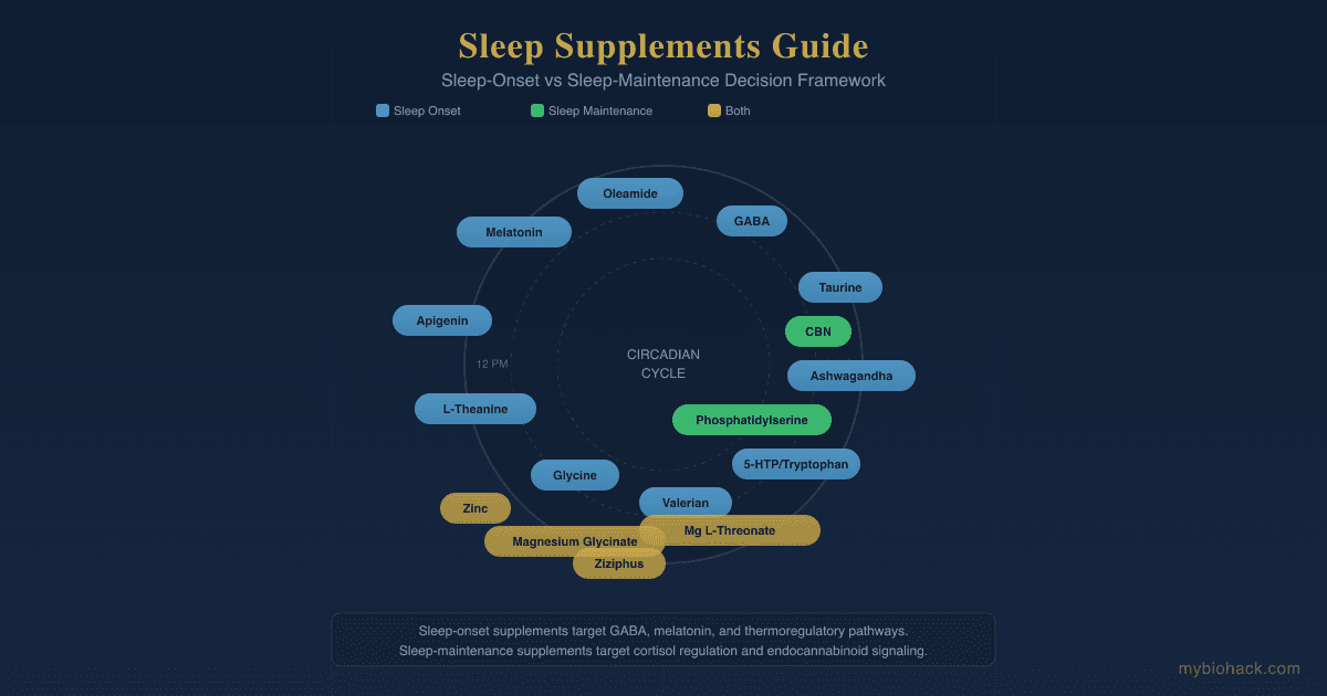
General sleep hygiene and supplementation (magnesium, melatonin timing, circadian light exposure) genuinely help sleep quality, and I write about this at length in my Sleep Supplements Guide and my own experiments in My Sleep Experiments And Favorite Sleep Hacks and How I Consistently Get 3-4 Hours Of Deep Sleep Every Night.
None of that fixes an anatomically or physiologically driven breathing problem, and I have seen clients spend years optimizing sleep hygiene while the actual driver (a narrow airway, a deviated septum, high loop gain) went untested and untreated the entire time.
Testing
Objective Sleep Testing
An in-lab polysomnogram with RERA scoring is the correct diagnostic test whenever UARS is suspected, since standard home testing is structurally blind to the events that define it, as covered in the diagnosis section above.
Cardiovascular And Metabolic Consequence Markers
Given how directly OSA drives endothelial dysfunction, insulin resistance, and inflammation, I use the Cardio Zoomer (Vibrant Wellness) to assess endothelial function, ApoB, lipid particle count, and inflammatory markers together, since these are the downstream consequences most likely to show up before a client notices any daytime symptoms.
I also use hs-CRP (Quest Diagnostics, via Fullscript) as an inexpensive standalone inflammation marker, and the Insulin Resistance Panel With Score (Quest Diagnostics, via Fullscript) to catch the metabolic consequences of intermittent hypoxia before fasting glucose alone would flag anything.
Cortisol And Autonomic Load
Chronic sympathetic activation from nightly apneic events disrupts normal cortisol rhythm, so I use the DUTCH Complete (Precision Analytical) or the Hormone Zoomer (Vibrant Wellness) to assess cortisol awakening response and full-day rhythm rather than relying on a single-point blood draw, which routinely misses this pattern.
Oxidative Stress And Mitochondrial Load
Because intermittent hypoxia generates reactive oxygen species similarly to ischemia-reperfusion injury, I use the Cellular Zoomer (Vibrant Wellness) to assess oxidative stress and mitochondrial markers in clients with confirmed or suspected sleep-disordered breathing.
For Overlapping POTS Or Dysautonomia
For clients where POTS or dysautonomia is already in the picture, the store's POTS testing bundle combines the Cellular, Hormone, Gut, and Neural Zoomers into one panel, which is the combination I reach for most often given how frequently I see sleep-disordered breathing and autonomic dysfunction overlap clinically.
Mechanisms Of Action
Simple:
- The upper airway is a muscular tube that naturally relaxes during sleep.
- If it narrows too far, either from anatomy, nasal resistance, or unstable breathing control, airflow becomes turbulent or blocks entirely.
- The brain briefly wakes up to reopen the airway, and that wake-up (not just the oxygen drop) is what fragments sleep and triggers the stress response.
Advanced:
- Pharyngeal collapsibility. The airway's tendency to close is governed by the balance between structural support (bone, soft tissue) and active neuromuscular dilator tone; during sleep, tone drops physiologically, and in an anatomically narrow airway this drop is enough to raise the critical closing pressure past the point of stability. R
- Loop gain instability. In a high-loop-gain airway, a small CO2 rise after a respiratory event triggers an exaggerated compensatory hyperventilation, which then overshoots, drops CO2 too far, and triggers a subsequent central apnea or hypopnea, creating a self-sustaining oscillation independent of anatomy alone. R
- Arousal threshold. A low arousal threshold means cortical arousal occurs before compensatory dilator muscle activation has time to reopen the airway on its own, so the airway never gets the chance to self-correct and sleep fragments on every event instead of only the severe ones. R
- Sympathetic and catecholamine surge. Each arousal event triggers a burst of norepinephrine and epinephrine release, producing acute spikes in heart rate and blood pressure that repeat dozens to hundreds of times nightly and compound over years into sustained hypertension risk. R
- Intermittent hypoxia and reoxygenation injury. Repeated cycles of desaturation followed by rapid reoxygenation generate reactive oxygen species through a mechanism analogous to ischemia-reperfusion injury, depleting antioxidant capacity and driving the endothelial and metabolic consequences described above. R
- Glymphatic clearance failure. Slow-wave sleep drives the majority of glymphatic clearance of interstitial waste through perivascular AQP4 channels, and sleep fragmentation from any cause (apnea, RERA, or arousal) truncates the deep-sleep window this clearance depends on. R
Genetics
Sleep apnea has a substantial heritable component independent of BMI, and genome-wide association studies have begun identifying specific risk loci, though the field is still earlier-stage than for many other chronic conditions.
TNF (Tumor Necrosis Factor Alpha)
TNF encodes tumor necrosis factor alpha, a pro-inflammatory cytokine central to the inflammatory response.
A functional promoter polymorphism in the TNF-alpha gene has been associated with excessive daytime sleepiness in children with OSA, independent of AHI itself, suggesting inflammatory genetic background can shape symptom severity beyond event count alone.
ADRB2 (Beta-2 Adrenergic Receptor)
ADRB2 encodes the beta-2 adrenergic receptor, which influences smooth muscle tone, including in tissue relevant to upper airway dilator function and autonomic responses during sleep.
Single nucleotide polymorphisms in ADRB2 have been associated with OSA severity in pediatric patients, consistent with a role in how the airway responds to the sympathetic surges described above.
APOE (Apolipoprotein E)
APOE encodes apolipoprotein E, and its epsilon-4 allele (APOE4) is the best-established common genetic risk factor for Alzheimer's disease.
The APOE4 allele appears more frequently in children with OSA than in unaffected children, and in adults, OSA severity is associated with greater amyloid-beta burden specifically in APOE4 carriers, which ties directly back to the glymphatic clearance mechanism described above.
More Research
- Cardiovascular risk beyond AHI. The American Heart Association's 2021 scientific statement and a 2024 JACC state-of-the-art review both place OSA as an independent, mechanistically distinct cardiovascular risk factor rather than a passive marker of obesity or aging. R R
- Hypoglossal nerve stimulation candidacy is narrowing, not widening. DISE-confirmed complete concentric collapse remains one of the strongest predictors of treatment failure, so the "no CPAP, get Inspire" framing skips a screening step that materially changes outcomes. R
- Myofunctional therapy evidence is real but not as strong as the frequently repeated 50%/62% statistic implies on its own. The Cochrane reviewers who re-examined the same evidence base called for larger trials before treating it as a standalone therapy, which is a genuine tension worth sitting with rather than resolving in either direction. R R
- Testing. For a general starting point before narrowing into the condition-specific panels above, I use the Foundation Zoomer (Vibrant Wellness) to establish a baseline before deciding which follow-up panels a client actually needs.
- Tirzepatide's cardiometabolic effects in OSA go beyond weight loss alone. A secondary analysis of the SURMOUNT-OSA trial reported cardiometabolic benefits that may not be fully explained by weight reduction, which is an open mechanistic question worth watching as more data accumulates. R
Jacob Gordon
INHC, FMT-C
Integrative Nutrition Health Coach
I cover mold illness, post-viral recovery, methylation, and complex chronic disease, drawing on ten years of clinical research, work inside a functional medicine clinic, and my own recovery from all of it. Every claim here is cited.
Book a ConsultationRelated Protocols & Supplements
Deep-dive chapters and recommended supplements for this topic
Electrolyte Complex
1 scoop/day
CoQ10
200mg/day
Magnesium Glycinate
400mg at bedtime