The Endothelial Health Protocol: A Complete Guide To Glycocalyx Repair, Vascular Function, And Reversing Endothelial Dysfunction
Back to Research Library
Vascular Health

The Endothelial Health Protocol: A Complete Guide To Glycocalyx Repair, Vascular Function, And Reversing Endothelial Dysfunction

|21 min read|0 likes

This article contains affiliate links. As an Amazon Associate, MyBioHack earns from qualifying purchases at no extra cost to you. We only link products we research and stand behind.

Endothelial dysfunction is the common thread linking cardiovascular disease, diabetes, long COVID, POTS, and most chronic inflammatory conditions, yet it remains undiagnosed in millions of people because conventional medicine does not look for it until a heart attack or stroke forces the issue.

In this post, we will discuss what endothelial dysfunction actually is, how it develops through glycocalyx degradation and the Junction Dysfunction framework, which conditions overlap with it, how to test for it, and a complete step-by-step protocol to restore vascular health.


Endothelial glycocalyx structure comparing the healthy state with thick heparan sulfate, hyaluronic acid, and chondroitin sulfate fibers to the degraded state with thin, broken fibers and exposed tight junctions

Basics Of Endothelial Dysfunction

The endothelium is a single-cell layer lining every blood vessel in the body, and it is the largest organ most people have never heard of.

It covers roughly 4,000 to 7,000 square meters of surface area in the average adult, the equivalent of a tennis court.

The endothelial surface is coated by the glycocalyx, a negatively charged, gel-like mesh of proteoglycans, glycosaminoglycans (GAGs), and glycoproteins that acts as the gatekeeper between blood and tissue.

When the glycocalyx is intact (see the Glycocalyx chapter for a deeper dive), the endothelium regulates vascular tone, prevents clotting, controls what passes into tissues, and keeps immune cells from sticking where they do not belong.

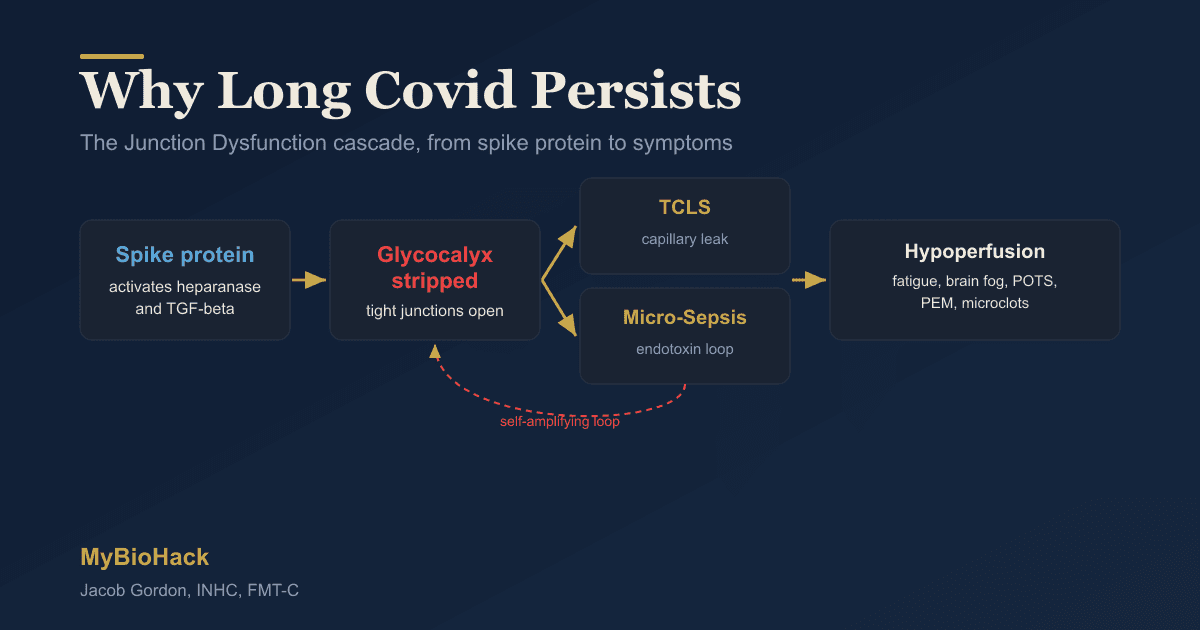
Endothelial dysfunction is the breakdown of these protective systems, and in my framework (the Junction Dysfunction (JD) model), it is driven by two interconnected subpathologies: Transient Capillary Leak Syndrome (TCLS) and Micro-Sepsis (MSS).

I coined the terms TCLS and MSS to describe the specific mechanisms that convert a healthy glycocalyx into a sieve.

TCLS is the microvascular version of capillary leak syndrome, where fluid and solutes escape the vasculature at the capillary level without registering on classical volume sensors. R

MSS is the chronic, sub-lethal version of sepsis triggered by LPS (lipopolysaccharide) auto-intoxication and constant TLR4 signaling, operating below the threshold of clinical sepsis diagnosis but using the same inflammatory machinery. R

The glycocalyx sits at the center of both pathologies (see The Interstitium for more on the fluid compartment directly affected by glycocalyx breakdown), and its degradation is the earliest detectable event in the progression toward endothelial dysfunction. R

What Causes Endothelial Dysfunction

Hyperglycemia And Insulin Resistance

Chronic high blood sugar alters the sulfation patterns of glycosaminoglycans in the glycocalyx and prevents hyaluronan from binding properly to the endothelial surface. R

Acute hyperglycemia alone causes measurable glycocalyx shedding, endothelial dysfunction, and coagulation activation within hours in healthy volunteers. R

The neuraminidase-ANO6-ADAM17 axis has recently been identified as a specific mechanism through which diabetes degrades the mechanosensor glypican-1 from the endothelial surface. R

Glycation And AGE/RAGE Signaling

Advanced glycation end products (AGEs) from high blood sugar and cooked foods bind the RAGE receptor on endothelial cells, activating NF-kB and triggering oxidative stress that sheds the glycocalyx. R

Soluble RAGE (sRAGE) fragments further amplify inflammation through a feed-forward loop that blocks nitric oxide signaling and opens tight junctions. R

Chronic Inflammation And Cytokine-Driven Shedding

TNF-alpha, IL-1 beta, and IL-6 activate matrix metalloproteinases (MMPs) and hyaluronidases that enzymatically cut through glycocalyx components. R

Heparanase specifically cleaves heparan sulfate from the glycocalyx, releasing GAG-bound cytokines that sustain local inflammation and drive further degradation in a self-reinforcing loop. R

Oxidative Stress And eNOS Uncoupling

Reactive oxygen species (ROS) directly destroy GAGs through oxidative cleavage, and they convert the normally protective endothelial nitric oxide synthase (eNOS) from a NO-producing enzyme into a superoxide-producing enzyme when its cofactor tetrahydrobiopterin (BH4) is depleted. R

This is called eNOS uncoupling, and it creates peroxynitrite (ONOO-), a highly reactive molecule that further damages the glycocalyx and endothelial cells. R

Shear Stress Loss And Hemodynamic Factors

The glycocalyx is a mechanosensor, meaning it requires appropriate shear stress from blood flow to maintain its structure.

Disturbed or reduced blood flow, as seen in sedentary behavior, atherosclerosis-prone arterial regions, and microcapillary loss, activates hyaluronidase and causes thinning of the glycocalyx. R

This is one reason that endothelial dysfunction preferentially develops at arterial bifurcations and curved segments where flow is turbulent. R

Infection And Post-Viral Mechanisms

SARS-CoV-2 spike protein directly binds heparan sulfate on the glycocalyx, and it activates TLR4 and RAGE signaling that causes rapid glycocalyx shedding. R

In long COVID, elevated ANGPT2 (angiopoietin-2) levels are consistent with ongoing glycocalyx loss and endothelial dysfunction that persists months after the acute infection clears. R

How The Endothelium Works (And How It Breaks)

The Glycocalyx As The First Line Of Defense

The glycocalyx is roughly 0.5 to 5 micrometers thick on most endothelial surfaces, and it is composed of three main structural classes.

Proteoglycans (syndecans and glypicans) anchor into the endothelial cell membrane and extend outward, carrying long GAG chains.

Glycosaminoglycans include heparan sulfate (the most abundant, about 50 to 90 percent of the glycocalyx), chondroitin sulfate, and hyaluronic acid (which is not attached to a core protein but binds directly to the CD44 receptor).

Glycoproteins include selectins, integrins, and other adhesion molecules embedded within the glycocalyx mesh.

The negative charge created by sulfated GAGs repels red blood cells and platelets, maintaining the zeta potential that keeps blood flowing and prevents inappropriate clotting. R

Mechanotransduction And Nitric Oxide Signaling

When blood flows across the glycocalyx, the shear force is transmitted through the glycocalyx structure to the endothelial cell surface.

This mechanical signal activates eNOS to produce nitric oxide (NO), which relaxes vascular smooth muscle and keeps arteries dilated. R

Without an intact glycocalyx, the endothelial cell cannot sense shear stress, eNOS does not activate, and the vessel loses its ability to dilate on demand. R

Endothelial health protocol ladder with four tiers: glycocalyx substrate support at the foundation, inflammation modulation, nitric oxide optimization, and structural support at the top
The endothelial health protocol organized in four ascending tiers. Start at the foundation with glycocalyx substrates and build up.

The Junction Dysfunction Cascade

In my framework, the sequence of events that converts a healthy endothelium into a dysfunctional one follows a predictable cascade.

PAMP/DAMP activation from infection, injury, or microbiome breach causes immune cells to release hyaluronidase, heparanase, and MMPs that cut through the glycocalyx.

Specific enzyme-substrate pairs involved include hyaluronidase cutting hyaluronic acid, heparanase cutting heparan sulfate, and MMPs cutting chondroitin sulfate.

ROS and RNS directly destroy GAGs through oxidative cleavage.

Once the glycocalyx is thinned, tight junctions between endothelial cells become exposed, and TCLS begins.

Fluid leaks from the intravascular space into the interstitium, creating intravascular hypovolemia and interstitial edema simultaneously. R

This fluid shift activates the renin-angiotensin-aldosterone system (RAAS) paradoxically, with high Angiotensin II despite inappropriately low renin and aldosterone, because classical volume sensors cannot detect microvascular fluid loss. R

This is my hypothesis for why POTS patients show this exact pattern of renin-aldosterone paradox.

MSS then develops as LPS from the gut (which enters circulation through the now-leaky gut endothelium, a process I call Endotoxin Looping) continuously activates TLR4 signaling, creating a state of chronic innate immune activation followed by immune paralysis. R

Endothelial Dysfunction And Overlapping Conditions

Cardiovascular Disease And Atherosclerosis

Endothelial dysfunction is the earliest detectable precursor to atherosclerosis, preceding plaque formation by years or decades.

Glycocalyx thinning occurs at arterial regions exposed to disturbed flow, and those are precisely the sites where atherosclerotic plaques later develop. R

Loss of the glycocalyx allows LDL particles to enter the vessel wall, where they become oxidized and trigger foam cell formation and plaque development. R

Long COVID And Post-Viral Syndromes

Persistent endothelial dysfunction is one of the most well-documented findings in long COVID, with elevated ANGPT2, syndecan-1, and hyaluronic acid levels indicating ongoing glycocalyx shedding. R

Endothelial function improved significantly with sulodexide treatment in long COVID patients, with corresponding reductions in chest pain and palpitations. R

Diabetes And Metabolic Syndrome

Both type 1 and type 2 diabetes cause measurable glycocalyx thinning, and the extent of thinning correlates with glycemic control and the presence of diabetic complications. R

Low molecular weight fucoidan has been shown to restore the diabetic endothelial glycocalyx by targeting neuraminidase-2 (NEU2), a specific enzyme upregulated by hyperglycemia. R

Hypertension

Hypertensive patients have reduced glycocalyx thickness, and the degree of thinning correlates with vascular stiffness and blood pressure levels. R

High sodium intake reduces heparan sulfate content in the glycocalyx and is associated with syndecan-1 shedding. R

POTS And Dysautonomia (VAD/ABVAD)

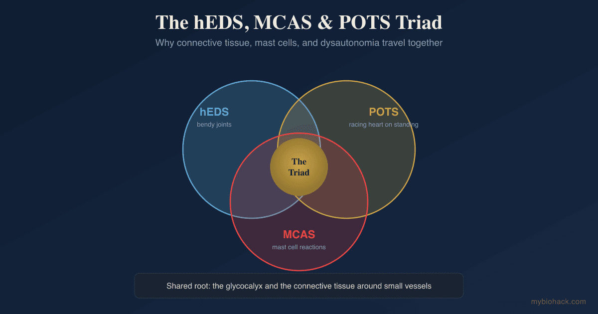
In my framework, Vaso-Adaptive Disorder (VAD, which replaces the term "Vascular POTS") and Adrenergic-Based Vaso-Adaptive Disorder (ABVAD, replacing "Hyperadrenergic POTS") are downstream consequences of TCLS-driven endothelial dysfunction.

When the glycocalyx is degraded and microcapillaries are lost, red blood cells shoot through fewer channels at higher velocity (force equals mass times acceleration), creating inappropriate tachycardia even when blood pressure is normal.

This is explained in detail in the Junction Dysfunction chapter on TCLS and the VAD chapter.

Mast Cell Activation Syndrome

Mast cells reside in connective tissue near blood vessels, and they degranulate in response to the hypoxia signals generated by microcapillary loss and TCLS.

Histamine from mast cells further opens tight junctions, creating a bidirectional worsening loop between endothelial dysfunction and mast cell activation. R

Autoimmune And Connective Tissue Disorders

Antibodies against endothelial cells, phospholipids, and clotting factors are commonly found in conditions like lupus, antiphospholipid syndrome, and rheumatoid arthritis.

I frame these antibodies not as attackers but as cleanup signals tagging damaged glycocalyx components and endothelial debris for removal (see my TGF-Beta1 post and the VEGF post for more on this frame).

How To Improve Endothelial Health

This protocol is organized into foundational, supplemental, and advanced tiers.

Start with the foundations, add supplements based on your testing and clinical picture, and escalate to advanced interventions if response is insufficient.

1. Restore Glycocalyx Substrates

The glycocalyx is built from specific sulfated polysaccharides, and providing the raw materials for its repair is the first step.

Glucosamine sulfate: Glucosamine is a direct precursor for hyaluronic acid and heparan sulfate synthesis, and it provides the amino sugar backbone needed for GAG chain formation. R

Chondroitin sulfate: Chondroitin sulfate is a major GAG component of the glycocalyx that provides structural integrity, and supplementation supports the replacement of chondroitin lost through MMP-mediated cleavage. R

Hyaluronic acid (HMW): High molecular weight hyaluronic acid (HMW-HA) provides anti-inflammatory signaling and scaffolding for glycocalyx repair, while low molecular weight HA is pro-inflammatory and should be avoided. R

2. Fucoidans (Sulfated Polysaccharides)

Fucoidans are sulfated polysaccharides extracted from brown seaweed that act as heparan sulfate mimetics, replacing the heparan that has been shed from the glycocalyx.

Both high molecular weight (HMW) and low molecular weight (LMW) fucoidans are needed for comprehensive glycocalyx support.

HMW fucoidans bind pathogenic bacteria and spike protein, activate NK cells, and provide neuroprotection.

LMW fucoidans cross cell membranes, promote vascular repair, and reduce endotoxemia.

Fucoidan (HMW): HMW fucoidan restores the endothelial glycocalyx by inhibiting heparanase and activating the ERK/MAPK and PI3K signaling pathways that drive glycocalyx synthesis. R

Low molecular weight fucoidan restores the diabetic endothelial glycocalyx by targeting neuraminidase-2 (NEU2), a key enzyme upregulated by hyperglycemia that cleaves sialic acid from the glycocalyx. R

Fucoidan selection guide comparing HMW and LMW fucoidans by molecular weight, source, primary mechanism, clinical applications, and signaling pathways
HMW and LMW fucoidans serve different but complementary roles in glycocalyx repair. Both are needed for comprehensive support.

Ecklonia cava: This brown seaweed contains phlorotannins that provide additional glycocalyx protection, and it is best taken at night because phlorotannins promote GABA activity and sleep. R

Products like Endocalyx Pro contain both fucoidan and glycocalyx substrate components and have shown the ability to increase glycocalyx thickness in human endothelial cells. R

3. Nitric Oxide Modulation (The Citrulline Path)

Jacob's clinical observation: Blind L-arginine supplementation is dangerous in the JD population because it reactivates latent viruses through the arginine-dependent replication cycle of herpesviruses, and without proper BH4 and SOD2 function, excess arginine feeds the peroxynitrite pathway.

L-citrulline: L-citrulline is converted to L-arginine in the kidneys and is the safer pathway for NO support. R

It does not directly activate viral replication (arginine is the rate-limiting substrate for viral protein synthesis, and citrulline bypasses this issue).

L-citrulline supplementation improves endothelial function, as measured by flow-mediated dilation (FMD), in patients with coronary artery disease. R

In hypertensive postmenopausal women, L-citrulline increased FMD and reduced aortic diastolic blood pressure. R

Combined citrulline and glutathione supplementation improves FMD and blunts blood pressure reactivity to stress. R

4. Pycnogenol (French Maritime Pine Bark Extract)

Pycnogenol: Pycnogenol is a standardized extract of Pinus pinaster bark (covered in detail in French Maritime Pine Bark) containing procyanidins, catechin, and taxifolin that augment endothelial-dependent vasodilation by increasing eNOS activity and NO production. R

In a double-blind, randomized, placebo-controlled crossover trial in patients with coronary artery disease, Pycnogenol (200 mg/day for 8 weeks) improved flow-mediated dilation from 5.3 to 7.0 percent and reduced oxidative stress measured by 15-F2t-isoprostane. R

For the methylation cycle support needed for BH4 recycling and glycocalyx repair, see the Methylation Complete Guide.

Pycnogenol also reduces endothelin-1 concentrations and supports healthy blood pressure in hypertensive patients. R

5. Boswellia Serrata (AKBA)

Boswellia serrata: The active compound acetyl-11-keto-beta-boswellic acid (AKBA) is a potent inhibitor of MMPs and 5-lipoxygenase, and it directly protects the glycocalyx by preventing the enzymatic cleavage of GAGs. R

AKBA prevents TNF-alpha-induced expression and activity of MMP-3, MMP-10, and MMP-12 in human microvascular endothelial cells. R

It also attenuates TGF-beta1-mediated vascular remodeling and reduces NF-kB activation in endothelial cells. R

Supercritical CO2-extracted boswellia provides the highest AKBA content, and this is the form I recommend.

6. Berberine

Berberine: Berberine is an alkaloid from Berberis species (detailed in my Berberine Benefits post) that improves endothelial function through multiple mechanisms.

It upregulates eNOS expression through AMPK activation, increases circulating endothelial progenitor cells, and reduces oxidative stress by downregulating NOX4 expression. R

Berberine upregulates endothelial progenitor cell number and function in healthy subjects through a mechanism related to NO production. R

In the JD Guide

Chapter 1

The Glycocalyx: The Root of It All

The glycocalyx is a microscopic gel layer coating every blood vessel in your body. When it breaks down, blood flow is impaired at the capillary level, the root mechanism behind Long COVID, POTS, MCAS, brain fog, and dozens of conditions conventional medicine treats as unrelated.

Pro members reading this now
Read it in Pro

It also prevents oxLDL-induced LOX1 expression and reduces the expression of adhesion molecules VCAM and ICAM. R

Berberine inhibits spike-ACE2 binding, which reduces ongoing endothelial damage in post-COVID contexts. R

7. Glycine And Collagen Support

Glycine: Glycine is the rate-limiting amino acid for collagen synthesis (collagen is about one-third glycine), and it also serves as a precursor for glutathione synthesis. R

Glycine improves endothelial function in aged animal models by enhancing eNOS expression and reducing superoxide and contractile prostanoids that blunt NO bioavailability. R

Collagen peptides: Hydrolyzed collagen provides glycine, proline, and hydroxyproline that support the structural integrity of the blood vessel wall, which is primarily composed of collagen type 3. R

8. Fasting And Ketosis

Intermittent fasting and nutritional ketosis create a repair window for the glycocalyx.

Beta-hydroxybutyrate (BHB) dampens MMP cleavage during fasting periods, reducing enzymatic glycocalyx shedding. R

Hyperglycemia prevents hyaluronan binding to the glycocalyx, so reducing blood glucose through fasting opens the window for repair.

The circadian protocol (morning light, time-restricted feeding, no late-night eating) supports the endothelial repair that occurs during overnight fasting periods.

9. Advanced Interventions

Sulodexide: Sulodexide is a purified mixture of heparan sulfate and dermatan sulfate that has been shown to improve glycocalyx dimensions and reduce vascular permeability in diabetic patients. R

In a study of 290 long COVID patients, sulodexide significantly improved endothelial function and reduced chest pain and palpitations. R

Endocalyx Pro: Endocalyx Pro is a dietary supplement containing fucoidan and glycocalyx substrate components that preserves the endothelial glycocalyx through ERK/MAPK signaling. R

In a pilot study in older adults, Endocalyx Pro improved FMD normalized to shear rate and increased capillary glycocalyx thickness in those not taking antihypertensives. R

IV Vitamin C: IV vitamin C replenishes BH4, supports eNOS coupling, and reduces oxidative load on the endothelium through hormetic H2O2 signaling. R

Hyperbaric oxygen therapy: HBOT (soft chamber, 1.2 to 1.5 ATA) supports HIF signaling and promotes angiogenesis in hypoxic tissues, helping rebuild microcapillary networks in conditions of established TCLS. R

EECP (Enhanced External Counterpulsation): EECP creates shear stress on the endothelium through external compression of the lower extremities, which stimulates eNOS activity and glycocalyx repair without requiring the patient to exercise. R

What To Stay Away From

High Glycemic Load Carbohydrates And Sugars

Hyperglycemia directly sheds the glycocalyx within hours of consumption.

Avoid refined sugars, fruit juices, high-glycemic grains, and any food that causes rapid blood glucose spikes.

Excess Sodium (In Processed Foods)

High sodium intake reduces heparan sulfate content in the glycocalyx and is associated with syndecan-1 shedding. R

L-Arginine Monotherapy

L-arginine supplementation in the absence of adequate BH4 and SOD2 function feeds the peroxynitrite pathway and creates more oxidative damage than benefit.

L-arginine also reactivates latent herpesviruses (HSV, EBV, CMV) because these viruses depend on arginine for replication.

Use L-citrulline instead, or ensure adequate BH4 status and SOD2 function before using arginine.

Excessive Alcohol

Alcohol directly damages the glycocalyx and increases vascular permeability, and it depletes the methylation cycle cofactors needed for glycocalyx repair.

Smoking And Vaping

Every inhalation of cigarette smoke delivers thousands of oxidants that directly destroy the glycocalyx, and nicotine (independent of smoke) causes vasoconstriction that reduces the shear stress needed for glycocalyx maintenance.

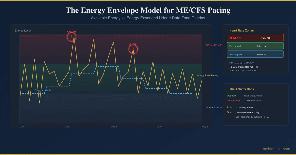
Overexercise (PEM)

Telling sick patients to exercise is counterproductive in the context of active glycocalyx degradation.

Post-exertional malaise is real and mechanistic, not deconditioning, and intense exercise during active glycocalyx degradation causes further shedding through shear-induced MMP activation.

Gentle movement (walking, recumbent cycling, very low intensity) is acceptable only when the patient is not in a post-exertional crash.

NSAIDs

Chronic NSAID use interferes with the endothelial repair mechanisms that depend on proper prostaglandin signaling through COX-2.

Testing

Imaging And Vascular Function Testing

Flow-mediated dilation (FMD) of the brachial artery using high-resolution ultrasound is the gold-standard non-invasive test for endothelial function, and it measures the artery's ability to dilate in response to shear stress. R

FMD is operator-dependent and technically demanding, so it is primarily used in research settings rather than routine clinical practice.

Peripheral arterial tonometry (finger plethysmography, EndoPAT) is a simpler non-invasive method that measures reactive hyperemia in the fingertip and correlates with endothelial function. R

Carotid intima-media thickness (CIMT) by ultrasound measures structural changes in the artery wall that reflect established endothelial damage and atherosclerotic progression. R

Blood And Urine Markers

Glycocalyx shedding markers: Elevated levels of syndecan-1, hyaluronic acid, and heparan sulfate in the blood indicate active glycocalyx degradation. R

I use the Cardio Zoomer (Vibrant Wellness) to assess comprehensive endothelial and cardiovascular markers, including lipid/lipoprotein profile, ApoB, insulin, metabolic markers, and ceramides.

ADMA (asymmetric dimethylarginine): ADMA is an endogenous inhibitor of eNOS that rises when the NO pathway is impaired, and elevated ADMA predicts cardiovascular events independently of traditional risk factors. R

hs-CRP: High-sensitivity C-reactive protein is a broad marker of systemic inflammation that correlates with endothelial dysfunction and cardiovascular risk.

ICAM-1, VCAM-1, E-selectin: These soluble adhesion molecules rise when the endothelium switches from a resting to an activated state, reflecting increased stickiness for leukocytes. R

Oxidized LDL (oxLDL) and LOX-1: Oxidized LDL binds the LOX-1 receptor on endothelial cells, triggering inflammatory signaling and glycocalyx shedding, and elevated levels indicate active oxidative modification of lipoproteins. R

ANGPT2 (Angiopoietin-2): ANGPT2 opposes ANGPT1 and destabilizes the endothelium, and elevated levels correlate with glycocalyx loss in long COVID and ME/CFS. R

VEGF and TGF-beta1: These growth factors are involved in maintaining vascular integrity, and their dysregulation is a hallmark of endothelial dysfunction (see my detailed posts on VEGF and TGF-beta1).

Functional Lab Panels

I use the Cellular Zoomer (Vibrant Wellness) to assess organic acids, mitochondrial function, and oxidative stress markers that provide indirect information about endothelial health.

I use the Toxin Zoomer (Vibrant Wellness) to assess mycotoxin and heavy metal burden, because toxicants directly damage the glycocalyx through oxidative stress.

I use the Long COVID Bundle (Vibrant Wellness) for post-viral patients, which combines the Cardio, Toxin, Gut, Cellular, and Viral zoomers into a comprehensive assessment.

I use the Nutrient Zoomer (Vibrant Wellness) to assess vitamins B6, B12, C, D, copper, zinc, and magnesium, because these are essential cofactors for eNOS function, SOD activity, and glycocalyx synthesis.

For nitric oxide pathway assessment, I use the fs-q-fasting-insulin and fs-q-insulin-resistance-panel (Quest via Fullscript) because insulin resistance is the most common driver of endothelial dysfunction in the general population.

Genetics Testing

I use the Methylation Genetics (Vibrant Wellness) or 3X4 Genetics test to assess the SNPs that determine individual glycocalyx repair capacity, eNOS function, and oxidative stress handling.

See the Genetics section below for the specific genes to evaluate.

Mechanisms Of Action

Simple:

  • The glycocalyx acts as a physical barrier that repels immune cells and platelets, preventing inappropriate inflammation and clotting.
  • When the glycocalyx is intact, shear stress from blood flow activates eNOS to produce NO, which keeps blood vessels dilated and flexible.
  • Sulfated polysaccharides from seaweed (fucoidans) mimic the structure of heparan sulfate and can directly incorporate into the glycocalyx or stimulate its repair.
  • Boswellia's AKBA inhibits the enzymes (MMPs) that cut the glycocalyx apart during inflammation.
  • Berberine activates AMPK, which increases eNOS expression and NO production while reducing oxidative stress.
  • Fasting and ketosis reduce blood glucose, which removes the hyperglycemia-driven block on glycocalyx repair and dampens MMP activity.
  • L-citrulline bypasses the liver's arginase barrier and converts to arginine in the kidneys, providing a safer route to NO production than direct arginine supplementation.

Advanced:

  • Heparanase Inhibition By Fucoidans: Fucoidans function as competitive substrates for heparanase, the enzyme that degrades heparan sulfate during inflammation. By acting as a heparan sulfate mimetic, fucoidan occupies the heparanase active site and prevents the enzyme from cleaving endogenous heparan sulfate from the glycocalyx. This preserves the glycocalyx's negative charge and its ability to regulate vascular permeability. R
  • Neuraminidase-2 (NEU2) Targeting By LMW Fucoidan: Low molecular weight fucoidan binds directly to neuraminidase-2 (NEU2) with a dissociation constant of approximately 3 microMolar, inhibiting its activity and downregulating its expression. NEU2 cleaves sialic acid from glycocalyx components, and its upregulation in hyperglycemia is a key driver of diabetic glycocalyx shedding. LMW fucoidan restores the glycocalyx by interrupting this neuraminidase-driven degradation pathway. R
  • ERK/MAPK And PI3K Pathway Activation: Fucoidan and glycocalyx-supporting supplements activate ERK/MAPK and PI3K signaling pathways in endothelial cells, which drive the synthesis and exocytosis of new glycocalyx components from pre-formed vesicles. Brefeldin A (a Golgi transport inhibitor) blocks this effect, indicating that the mechanism involves vesicular transport and exocytosis rather than direct incorporation of supplement components into the glycocalyx. R
  • Pycnogenol eNOS Upregulation: The procyanidins and bioflavonoids in Pycnogenol (catechin, taxifolin) increase eNOS expression and subsequent NO release from endothelial cells. This effect is completely abolished by the NOS inhibitor L-NMMA, confirming it is NO-dependent. Pycnogenol also reduces endothelin-1, a potent vasoconstrictor that is elevated in endothelial dysfunction. R
  • AKBA Inhibition Of MMPs And NF-kB: Acetyl-11-keto-beta-boswellic acid prevents TNF-alpha-induced expression of matrix metalloproteinases (MMP-3, MMP-10, MMP-12) in human microvascular endothelial cells. It also inhibits the IkB kinase complex, preventing NF-kB nuclear translocation and reducing expression of downstream inflammatory genes including MCP-1, IL-1 beta, VEGF, and tissue factor. This dual action protects the glycocalyx from both direct enzymatic cleavage and cytokine-driven shedding. R
  • Berberine AMPK-eNOS-NOX4 Axis: Berberine activates AMPK, which phosphorylates and activates eNOS at Ser1177, increasing NO production. It simultaneously downregulates NOX4, the NADPH oxidase isoform that generates superoxide in the endothelium. This dual mechanism improves NO bioavailability by increasing NO production and reducing superoxide-mediated NO destruction. Berberine also inhibits LOX-1 expression, reducing oxLDL uptake into endothelial cells and the subsequent inflammatory signaling. R
  • ADAM17-Glypican-1 Mechanosensor Protection: Hyperglycemia activates neuraminidase, which increases intracellular calcium, which activates anoctamin-6 (ANO6), which flips phosphatidylserine to the outer membrane leaflet, which activates ADAM17, which sheds glypican-1 (the mechanosensor) from the glycocalyx. This cascade is a newly identified mechanism of diabetic endothelial dysfunction. ANO6 or ADAM17 inhibition prevents this shedding. LMW fucoidan partially interrupts this cascade by inhibiting NEU2 upstream. R

Genetics

NOS3 (eNOS)

The NOS3 gene encodes endothelial nitric oxide synthase, the enzyme responsible for producing NO from L-arginine in the endothelium.

Polymorphisms in NOS3 reduce eNOS expression and activity, and they partially explain variability in cardiovascular drug responses and endothelial function. R

rs1799983 (Glu298Asp): This missense variant produces an eNOS protein that is more susceptible to proteolytic cleavage, reducing NO production capacity and increasing cardiovascular risk. R

rs2070744 (-786T>C): This promoter variant reduces NOS3 transcription by approximately 50 percent in the C allele, leading to lower baseline NO production and higher risk for hypertension and coronary artery disease. R

SOD2

The SOD2 gene encodes mitochondrial superoxide dismutase, the primary enzyme that quenches superoxide produced by the mitochondrial electron transport chain.

Jacob's clinical observation: Most clients with redox imbalances have SOD2 mutations at rs4880 (Ala16Val), which reduces SOD2 efficiency and increases the risk of eNOS uncoupling because excess superoxide converts NO to peroxynitrite.

rs4880 (Ala16Val): The Val allele reduces SOD2 import into the mitochondria by approximately 30 to 40 percent, increasing mitochondrial oxidative stress and the likelihood of peroxynitrite formation in the endothelium. R

AGER (RAGE)

The AGER gene encodes the receptor for advanced glycation end products (RAGE), which is activated by AGEs, S100 proteins, HMGB1, and the SARS-CoV-2 spike protein.

RAGE activation on endothelial cells triggers NF-kB signaling, glycocalyx shedding, and tight junction opening. R

rs2070600 (Gly82Ser): This variant alters RAGE binding affinity, and it may influence the inflammatory set point and susceptibility to glycocalyx damage in response to AGEs and spike protein. R

ACE2

The ACE2 gene encodes angiotensin-converting enzyme 2, which counter-regulates the RAAS by converting Angiotensin II to Angiotensin (1-7), a vasodilatory and anti-inflammatory peptide.

Lower ACE2 expression leaves Angiotensin II unopposed, driving AT1R-mediated vasoconstriction, inflammation, and glycocalyx shedding. R

rs2285666: This intronic variant affects ACE2 expression levels, with the GG genotype associated with higher ACE2 expression and potentially more favorable endothelial outcomes. R

SUOX

The SUOX gene encodes sulfite oxidase, which converts sulfite to sulfate for incorporation into heparan sulfate, chondroitin sulfate, and other sulfated GAGs.

Impaired sulfite oxidase function reduces the availability of sulfate for glycocalyx synthesis, limiting repair capacity.

rs7297662: Variants in SUOX slow sulfite-to-sulfate conversion, and this is clinically relevant for glycocalyx repair because a sulfur-deficient glycocalyx has reduced negative charge and is more easily shed. R

IFITM3

The IFITM3 gene encodes interferon-induced transmembrane protein 3, which restricts viral entry into cells by altering membrane fluidity.

rs12252 (CC genotype): This variant is associated with more severe COVID-19 outcomes, likely because reduced IFITM3 function allows greater viral entry and replication, which in turn causes more endothelial damage and glycocalyx shedding. R

More Research

  • Endothelial progenitor cells (EPCs) as a therapeutic target: Circulating EPCs home to sites of endothelial damage and participate in vascular repair. Berberine and exercise both increase EPC mobilization, and the degree of EPC mobilization correlates with FMD improvement. EPC count and function may become a clinically useful biomarker for endothelial repair capacity. R
  • Extracellular microvesicles (EMVs) as biomarkers: Endothelial-derived microvesicles are shed from damaged endothelial cells and carry surface markers (CD31, CD62E, CD144) that reflect the state of the parent cell. Their levels correlate with endothelial function and decline when vascular health improves. EMVs may become the preferred clinical test for endothelial dysfunction because they are stable in frozen plasma and less operator-dependent than FMD. R
  • The circadian timing of glycocalyx repair: Glycocalyx synthesis follows a circadian rhythm, with peak repair occurring during overnight fasting and sleep. Disruption of this rhythm (night eating, shift work, blue light at night) impairs glycocalyx maintenance. The circadian protocol I describe in the JD guide is not optional for restoring endothelial health.
  • Glycocalyx measurement by GlycoCheck: The GlycoCheck system uses sidestream dark-field imaging of sublingual microvessels to estimate glycocalyx thickness by measuring the perfused boundary region (PBR). This may become the most practical clinical tool for assessing glycocalyx status, though standardized protocols and reference ranges are still being developed. R
  • For biomarker testing I use the Cardio Zoomer to assess endothelial health comprehensively, and I combine it with the Cellular Zoomer to evaluate the oxidative stress and mitochondrial function that directly impact eNOS coupling and glycocalyx integrity.
  • For readers who want to explore the mechanistic detail behind glycocalyx repair, the Improving The Glycocalyx chapter is the most comprehensive resource I have written on this topic, and it covers the full protocol with dosing, timing, and caveats that go beyond what this post can include.
  • Jacob's hypothesis: The bidirectional ANGPT/TIE2-glycocalyx loop may explain why some patients respond rapidly to glycocalyx-supporting therapies while others do not. When the glycocalyx is degraded, ANGPT/TIE2 destabilizes, causing more glycocalyx shedding via heparanase release, creating a self-reinforcing loop. Breaking this loop may require simultaneous glycocalyx substrate support AND ANGPT/TIE2 stabilization (through direct ANGPT1 agonism or TIE2 activation), though specific therapeutics for this are still under investigation.
  • If your endothelial dysfunction is driven by post-viral mechanisms, the Long COVID Natural Treatment Protocol covers the immune and viral clearance aspects that must be addressed alongside the glycocalyx repair strategies in this post.
JG

Jacob Gordon

INHC, FMT-C

Board Certified Health Coach

I spent years battling unexplained chronic illness before discovering biohacking, epigenetics, and functional medicine. Now I share that research at MyBioHack to help others find their own answers.

Book a Consultation

Related Protocols & Supplements

Deep-dive chapters and recommended supplements for this topic

Recommended Supplements

Electrolyte Complex

1 scoop/day

CoQ10

200mg/day

Magnesium Glycinate

400mg at bedtime

Protocols from Jacob's Junction Dysfunction guideView Full Guide

What's Working For You?

What has been your experience with this topic? Unlock the full community archive with Pro.

Sign in to share your stack