TGF-Beta1: Why You Should Not Reflexively Try To Lower It
By Jacob Gordon, INHC, FMT-CThis article contains affiliate links. As an Amazon Associate, MyBioHack earns from qualifying purchases at no extra cost to you. We only link products we research and stand behind.
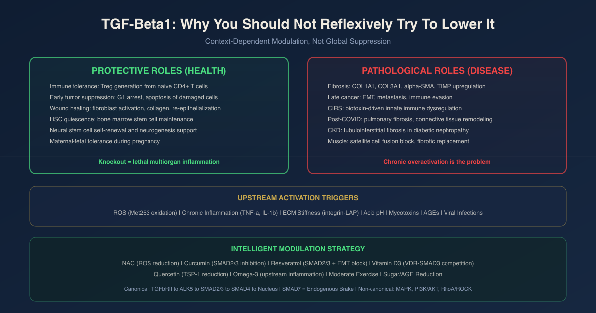
TGF-beta1 (transforming growth factor beta 1, encoded by the TGFB1 gene) is one of the most context-dependent molecules in human biology, simultaneously a tumor suppressor, an immune tolerance enforcer, a wound healer, and, when chronically elevated or dysregulated, a primary driver of fibrosis and cancer metastasis.
In this post, we will discuss what TGF-beta1 actually is, why eliminating it is catastrophically dangerous, what it does in health, what goes wrong when it becomes chronically elevated, which conditions involve TGF-beta1 dysregulation, how to modulate it intelligently rather than suppress it bluntly, and what the genetics look like.
What TGF-Beta1 Is
TGF-beta1 is a 25 kDa homodimeric cytokine and the most studied member of the transforming growth factor beta (TGF-beta) superfamily, which includes over 30 structurally related proteins including bone morphogenetic proteins (BMPs), activins, and nodal.
It is encoded by the TGFB1 gene on chromosome 19q13. R
It is expressed by virtually every cell type in the human body and signals through receptors found on virtually every cell type.
This ubiquity is not accidental.
TGF-beta1 functions as a master coordinator of tissue integrity, coordinating immune surveillance, growth arrest, ECM maintenance, and injury repair across the whole organism. R00851-6)
The three isoforms:
TGF-beta1, TGF-beta2, and TGF-beta3 share 70 to 82% amino acid homology and signal through the same receptors, but are not interchangeable in vivo.
TGF-beta1 is the predominant isoform in the immune system and is the most abundant and active isoform in most disease contexts.
TGF-beta2 knockout mice show a wide range of developmental defects in heart, lungs, bones, eyes, and craniofacial structures.
TGF-beta3 knockout mice die shortly after birth due to cleft palate and abnormal lung development.
Each isoform has distinct tissue-specific roles, which is important context for understanding why blunt TGF-beta inhibition carries significant risks. R
Latency: most TGF-beta1 is not active:
TGF-beta1 is synthesized as a pre-pro-peptide and secreted in a latent complex in which the mature cytokine is noncovalently associated with its own propeptide, called the latency-associated peptide (LAP).
In this latent form, TGF-beta1 cannot bind its receptors.
Only a small fraction of total tissue TGF-beta1 is biologically active at any given time.
The LAP must be displaced before signaling can occur.
Activation mechanisms include:
- Integrin-mediated activation: integrins alphaVbeta6 (epithelial) and alphaVbeta8 pull on LAP via cytoskeletal force, causing conformational change and active TGF-beta1 release (the most physiologically established mechanism in vivo) R
- Thrombospondin-1 (TSP-1): binds to LAP at a specific sequence and displaces it non-proteolytically; is a major physiological activator in vivo R
- Reactive oxygen species (ROS): oxidize a specific methionine residue (Met253) on TGF-beta1 LAP, causing conformational change and activation; the only known isoform-specific ROS activation mechanism R
- Proteases: matrix metalloproteinases (MMP-2, MMP-9), plasmin, and other enzymes can cleave LAP; relevant in injury and inflammation R
- Acidification: low pH (as occurs in tumor microenvironments and osteoclast resorption lacunae) directly activates latent TGF-beta1 R
This latency system is a key regulatory checkpoint.
Reducing total TGF-beta1 expression and reducing TGF-beta1 activation are mechanistically distinct targets with different risk profiles.
Why You Do Not Want To Fully Suppress TGF-Beta1
This is the section most health writers omit, and it is the most important one to understand before doing anything to modulate TGF-beta1.
In 1992, two independent research groups reported that mice with global knockout of the Tgfb1 gene died within the first few weeks of life.
The cause of death was not cancer or fibrosis.
It was massive, uncontrolled, multiorgan inflammation.
The heart, lungs, liver, and salivary glands were infiltrated with inflammatory cells.
Without TGF-beta1, the immune system could not regulate itself, and it destroyed its own host. R
T cell-specific deletion of the TGF-beta receptor (TGFbetaRII) in mice produces the same lethal inflammatory phenotype, confirming that TGF-beta1 signaling in T cells specifically is required for immune tolerance and survival. R
What happens without TGF-beta1:
Without TGF-beta1, there are no functional regulatory T cells (Tregs) in the periphery.
Tregs are the primary immune cell population that prevents autoimmune attack, dampens excessive inflammation, and maintains tolerance to self-antigens.
TGF-beta1 is required to generate and maintain peripheral Tregs from conventional T cells.
Remove TGF-beta1 signaling and Tregs collapse.
Remove Tregs and the immune system attacks every organ it can reach. R
Deficient TGF-beta1 in disease:
In type 1 diabetes-prone NOD mice, Treg function deteriorates as TGF-beta1-producing Tregs decrease with age.
The decline of TGF-beta1 production by Tregs directly correlates with disease onset and progression.
Restoring TGF-beta1 signaling in the islets of these mice suppresses diabetes development. R
In human autoimmune diseases, defects in TGF-beta1 expression or signaling are associated with the onset of systemic lupus erythematosus, rheumatoid arthritis, and inflammatory bowel disease. R
Chronic skin wounds fail to heal partly because TGF-beta1 signaling is insufficient to drive granulation tissue formation and re-epithelialization. R
In Alzheimer's disease, reduced TGF-beta1 signaling correlates with impaired neural stem cell self-renewal and may contribute to progressive neurodegeneration. R
The clinical problem with blanket TGF-beta inhibition:
As of 2023, 124 TGF-beta blocking agents had been identified, only two received regulatory approval (both in cancer), and 73 remained in clinical investigation.
The fundamental challenge is cardiovascular toxicity: global TGF-beta inhibition causes cardiac valve abnormalities, inflammatory lesions, and other off-target effects in animal models.
Phase II and III trials of pan-TGF-beta inhibitors have been complicated by this toxicity profile. R
The takeaway for functional medicine:
Aggressively suppressing TGF-beta1 with high doses of multiple inhibitory compounds simultaneously, particularly in someone with existing autoimmune tendencies or a history of chronic infection, is not a safe strategy.
The goal in most chronic illness contexts is not to eliminate TGF-beta1 but to normalize it: reduce chronic overactivation in specific pathological contexts (fibrosis, tumor promotion, chronic mold illness) while preserving its essential physiological functions.
What TGF-Beta1 Does In Health
Understanding what TGF-beta1 is protecting before attempting to modulate it is prerequisite to doing so safely.
Immune tolerance:
TGF-beta1 is the primary driver of peripheral regulatory T cell (Treg) induction from naive CD4+ T cells.
It suppresses activation-induced T cell proliferation.
It dampens Th1 and Th2 effector function.
It prevents autoimmune attack of self-tissues by keeping the immune system in check after an antigen has been cleared. R
Wound healing and tissue repair:
TGF-beta1 is the central orchestrator of acute wound healing.
After injury, platelets release TGF-beta1 at the wound site, initiating inflammation and recruiting immune cells.
TGF-beta1 then activates fibroblasts to produce collagen and form granulation tissue.
It drives re-epithelialization by stimulating keratinocyte migration.
Fibroblast-derived TGF-beta1 is specifically required for proper vascularization of early wounds: mice with fibroblast-specific Tgfb1 deletion show hemorrhage and impaired vascular density in healing wounds. R
Tumor suppression in early cancer:
TGF-beta1 suppresses early tumor development by arresting cell division at the G1 checkpoint, inducing apoptosis, preventing excessive epithelial proliferation, and suppressing excessive inflammation that could promote mutagenesis. R
This early tumor suppressor role is the most underappreciated aspect of TGF-beta biology.
Skeletal muscle satellite cell regulation:
TGF-beta1 regulates muscle repair by modulating satellite cell (muscle stem cell) activation.
It participates in the switch between the inflammatory phase of muscle injury and the regenerative phase.
At physiological levels, it coordinates the timing of muscle regeneration.
At pathologically elevated levels, it inhibits myoblast fusion, drives fibrosis, and impairs muscle regeneration. R
Hematopoiesis and bone marrow homeostasis:
TGF-beta1 maintains hematopoietic stem cell quiescence in the bone marrow niche.
It is required for normal erythropoiesis.
Dysregulation of TGF-beta signaling in hematopoiesis contributes to myelodysplastic syndrome, anemia of chronic disease, and aplastic anemia. R
Neural stem cell maintenance:
TGF-beta1 supports the self-renewal and maintenance of neural stem cells.
In the diseased brain, amyloid-beta and tau pathology interfere with TGF-beta1 signaling, potentially impairing neurogenesis and contributing to progressive neurodegeneration. R
Maternal-fetal tolerance:
TGF-beta1 produced by peripheral Tregs (induced at sites colonized by maternal microbiota) is required for maternal tolerance of the allogeneic fetus.
CNS1-deficient female mice (unable to generate peripheral Tregs) exhibit increased fetal resorption when bred to allogeneic males. R
The Dual Role: Tumor Suppressor Turned Promoter
TGF-beta1's behavior in cancer is the clearest example of why context determines everything about this molecule.
Early cancer: TGF-beta1 as tumor suppressor
In normal epithelium and during early carcinogenesis, TGF-beta1 arrests the cell cycle at G1 via upregulation of cyclin-dependent kinase inhibitors (p21, p15, p27).
It promotes apoptosis of damaged or dysplastic cells.
It suppresses inflammatory environments that would otherwise favor mutagenesis.
Losing TGF-beta signaling, or losing downstream SMAD components, is a common early step in colorectal cancer, pancreatic cancer, and other epithelial cancers. R
Late cancer: TGF-beta1 as tumor promoter
Once a tumor has acquired the ability to evade or exploit TGF-beta1 signaling (typically by losing expression of SMAD4, gaining oncogenic RAS mutations, or reprogramming the TGF-beta response), TGF-beta1 reverses its role.
It now drives epithelial-to-mesenchymal transition (EMT): the process by which cancer cells lose their epithelial identity, acquire mesenchymal properties, detach from the primary tumor, and become invasive and metastatic. R
TGF-beta1 from the tumor simultaneously suppresses the antitumor immune response by promoting Treg accumulation in the tumor microenvironment, polarizing macrophages toward the M2 immunosuppressive phenotype, and inhibiting NK cell, CD8+ T cell, and dendritic cell activity. R
It also activates cancer-associated fibroblasts (CAFs) that produce ECM, promote angiogenesis, and create a structural scaffold that shields tumor cells from immune attack and drug delivery. R
The clinical implication:
Whether TGF-beta1 should be stimulated or suppressed in a cancer patient depends entirely on the cancer type, stage, and TGF-beta signaling status of the tumor.
In early-stage, SMAD-intact cancers, supporting TGF-beta1 tumor suppressor function makes conceptual sense.
In late-stage, SMAD-deficient cancers with high TGF-beta expression, inhibiting TGF-beta1 is the correct direction.
There is no blanket answer for cancer.
What Drives Chronic TGF-Beta1 Elevation
Chronic activation of TGF-beta1 is the problem, not the existence of TGF-beta1 itself.
Reactive oxygen species (ROS):
Chronic oxidative stress is one of the most potent and sustained activators of latent TGF-beta1, via direct oxidation of Met253 in LAP.
Mitochondrial dysfunction, chronic infection, smoking, environmental toxins, and metabolic syndrome all generate excess ROS that continuously activate TGF-beta1 from its latent pool. R
Chronic inflammation:
Inflammatory mediators including TNF-alpha, IL-1beta, and platelet-derived growth factor (PDGF) upregulate TGF-beta1 expression in fibroblasts and macrophages.
Additionally, the inflammatory microenvironment activates TSP-1 production, which drives further TGF-beta1 activation from its latent pool, creating a self-reinforcing loop. R
Tissue injury and ECM stiffness:
Mechanical forces: myofibroblasts in fibrotic tissue contract against stiff ECM, and this mechanical tension applied to the integrin-LAP-TGF-beta1 complex continuously activates TGF-beta1 even in the absence of inflammatory triggers.
Fibrotic ECM stiffness drives TGF-beta1 activation that drives more fibrosis in a self-perpetuating mechanical loop. R
Acid microenvironments:
Low-pH microenvironments, including tumor interstitium and areas of metabolic acidosis, directly activate latent TGF-beta1.
Physiological concentrations of lactic acid (produced in ischemic or hypoxic tissue) have been shown sufficient to activate TGF-beta1 in lung fibrosis models. R
Mold toxins (mycotoxins) and biotoxins:
Many mycotoxins stimulate TGF-beta1 production in airway epithelial cells and macrophages.
In CIRS (Chronic Inflammatory Response Syndrome) from water-damaged buildings, persistently elevated TGF-beta1 is one of the measurable markers of ongoing innate immune activation and is associated with the fibrotic and neurological components of biotoxin illness. R
Viral infections:
Multiple chronic viral infections upregulate TGF-beta1, including EBV-associated nasopharyngeal carcinoma, hepatitis B and C (driving liver fibrosis), and post-COVID-19 pulmonary fibrosis. R
Advanced glycation end products (AGEs):
AGEs, elevated in chronic hyperglycemia and aging, stimulate TGF-beta1 production in fibroblasts and mesangial cells, contributing to diabetic nephropathy, retinopathy, and generalized tissue fibrosis. R
TGF-Beta1 And Overlapping Conditions
Fibrotic conditions: TGF-beta1 is the master driver of fibrosis in essentially every organ.
The SMAD2/3 pathway downstream of TGF-beta1 activation directly upregulates collagen (COL1A1, COL3A1), fibronectin, alpha-smooth muscle actin (alpha-SMA), and tissue inhibitors of metalloproteinases (TIMPs), while simultaneously downregulating matrix metalloproteinases that would degrade excess ECM.
This combination produces net ECM accumulation and progressive organ stiffening in pulmonary fibrosis, hepatic fibrosis, cardiac fibrosis, renal fibrosis, and scleroderma. R
CIRS and biotoxin illness: Elevated serum TGF-beta1 is a recognized biomarker of CIRS, along with elevated MSH, low VIP, and abnormal complement activation patterns.
TGF-beta1 elevation in this context is believed to reflect a dysregulated innate immune response to persistent biotoxin exposure, with TGF-beta1 contributing to the interstitial fibrosis, neurological symptoms, and immune dysregulation characteristic of the condition. R
Autoimmune diseases: While TGF-beta1 deficiency causes autoimmunity in animal models, chronic tissue-specific TGF-beta1 elevation paradoxically contributes to the fibrotic tissue destruction seen in scleroderma, Sjögren's syndrome, and the joint destruction of advanced rheumatoid arthritis.
The immune tolerance role and the fibrotic role coexist and can be simultaneously active in the same individual. R
Cancer (late stage): Elevated serum TGF-beta1 in advanced solid tumors is associated with worse prognosis, higher metastatic potential, and resistance to immunotherapy in multiple tumor types including colorectal, pancreatic, breast, and non-small cell lung cancer. R
Post-COVID and post-viral syndromes: SARS-CoV-2 infection triggers TGF-beta1 elevation through ROS generation, inflammatory cytokine production, and direct viral induction in lung epithelial cells.
Persistent TGF-beta1 elevation post-COVID is mechanistically linked to the pulmonary fibrosis, connective tissue remodeling, and potentially to the interstitial fluid dysfunction seen in long COVID. R
Chronic kidney disease (CKD): Urinary TGF-beta1 is elevated in CKD and positively correlates with degree of renal fibrosis.
TGF-beta1 is the dominant driver of tubulointerstitial fibrosis in diabetic nephropathy, hypertensive nephropathy, and post-obstructive injury. R
Duchenne muscular dystrophy and muscle diseases: Chronically elevated TGF-beta1 in dystrophic muscle inhibits satellite cell fusion (blocking regeneration) while driving fibroblast activation and ECM accumulation, progressively replacing functional muscle with fibrotic scar tissue. R
How To Modulate TGF-Beta1 Intelligently
The strategic goal is context-specific modulation, not global suppression.
For most people with chronic illness involving elevated TGF-beta1, the target is reducing the upstream drivers of TGF-beta1 overactivation (ROS, chronic inflammation, ECM stiffness, biotoxin burden) while preserving the essential physiological functions.
1. Address The Upstream Drivers First
TGF-beta1 elevation is almost always a downstream consequence of something else: oxidative stress, persistent infection, biotoxin load, metabolic dysfunction, or mechanical tissue damage.
Treating it as the primary problem while leaving its drivers intact will produce insufficient and temporary results.
N-acetylcysteine (NAC): replenishes glutathione and directly reduces ROS that activate latent TGF-beta1 via the Met253 oxidation mechanism.
In multiple animal models of pulmonary fibrosis, NAC reduces TGF-beta1-mediated fibrosis by limiting ROS-dependent TGF-beta1 activation.
Typical dosing: 600 to 1200 mg daily. R
Alpha lipoic acid (ALA): a mitochondria-targeted antioxidant that inhibits hepatic stellate cell activation and TGF-beta1-induced fibrotic signaling in liver models; also reduces TGF-beta1 expression in peritoneal adhesion models.
Prefer the R-form.
Typical dosing: 300 mg daily. R
2. Curcumin
Curcumin (bioavailable form): the most evidence-backed natural compound for modulating TGF-beta1 signaling.
Curcumin inhibits TGF-beta1 signaling through multiple mechanisms: it reduces TGF-beta receptor 1 (TGFbetaR1) expression, inhibits SMAD2 and SMAD3 phosphorylation, suppresses fibroblast migration and proliferation, and reduces myofibroblast differentiation.
Anti-fibrotic effects of curcumin mediated through TGF-beta downregulation have been shown across hepatic, pulmonary, renal, and dermal fibrosis models. R
Importantly, curcumin's effect on TGF-beta is context-dependent: it can actually upregulate TGF-beta1 in early-stage cancer contexts where TGF-beta1 functions as a tumor suppressor.
This is the right direction.
It is not a blunt TGF-beta suppressor. R
Bioavailability is the primary limitation of curcumin.
Standard curcumin supplements have poor oral bioavailability.
Use phytosome forms (Meriva), nanoparticle formulations, or curcumin combined with piperine.
Typical dosing: 500 to 1500 mg phytosome curcumin daily with food.
3. Resveratrol
Resveratrol: inhibits TGF-beta1/SMAD signaling and blocks EMT driven by TGF-beta1 in multiple cancer and fibrosis models.
Resveratrol directly suppresses SMAD2/3 phosphorylation and nuclear translocation in fibrotic fibroblasts.
It also promotes the degradation of TGF-beta1-induced myofibroblast markers (alpha-SMA) and reduces collagen deposition in liver fibrosis models. R
Typical dosing: 250 to 500 mg daily. R
4. Vitamin D3
Vitamin D3: has documented anti-fibrotic activity via inhibition of TGF-beta1/SMAD3 signaling.
The vitamin D receptor (VDR) when activated by 1,25-dihydroxyvitamin D3 forms a complex with SMAD3 that competes with SMAD4, disrupting TGF-beta1-driven gene transcription.
Epidemiological associations between vitamin D deficiency and elevated fibrotic disease risk are consistent with this mechanism. R
Typical dosing: 5000 IU daily with K2; adjust based on serum 25(OH)D (target 50 to 80 ng/mL).
5. Quercetin
Quercetin: inhibits TGF-beta1-induced SMAD2/3 phosphorylation and reduces TGF-beta1 expression in fibrotic models.
Also acts upstream by reducing TSP-1 production (one of the major TGF-beta1 activators), reducing integrin alphavbeta6 expression, and generally dampening the inflammatory environment that drives TGF-beta1 upregulation. R
Typical dosing: 500 to 1000 mg daily.
6. Omega-3 Fatty Acids (EPA/DHA)
Fish oil (high EPA/DHA): EPA and DHA reduce inflammatory cytokines (TNF-alpha, IL-1beta) that drive TGF-beta1 upregulation.
They also reduce TGF-beta1 expression in lung fibrosis models and can modulate the lipid mediator environment to favor resolution of inflammation over TGF-beta1-mediated fibrosis. R
Typical dosing: 2 to 4 g combined EPA+DHA daily.
7. Aerobic Exercise (Moderate Intensity)
Moderate aerobic exercise reduces serum TGF-beta1 levels, reduces systemic oxidative stress (which would otherwise activate latent TGF-beta1), and improves insulin sensitivity (which reduces AGE formation).
High-intensity exercise without adequate recovery can temporarily spike TGF-beta1 as part of the acute tissue repair response, which is physiologically appropriate.
The goal is sustained moderate activity rather than episodic intense bursts.
8. Dietary Sugar And Processed Carbohydrate Reduction
Reducing glycemic load directly reduces AGE formation.
AGEs bind to their receptor (RAGE) on fibroblasts and mesangial cells, stimulating TGF-beta1 production.
This is the primary mechanism by which chronic hyperglycemia drives TGF-beta1-mediated fibrosis in diabetic complications.
In practice, reducing dietary sugar and refined carbohydrates is one of the most accessible upstream interventions available. R
9. Pirfenidone And Nintedanib (Prescription Context)
These are the two FDA-approved anti-fibrotic drugs referenced here for completeness, not as self-treatment options.
Pirfenidone reduces TGF-beta1 expression and downstream SMAD signaling and is approved for idiopathic pulmonary fibrosis (IPF).
Nintedanib is a multikinase inhibitor that blocks multiple pathways including PDGF, VEGF, and FGF signaling that intersects with TGF-beta1 in fibrotic loops, also approved for IPF.
If you have a diagnosed fibrotic condition, these drugs are the most evidence-backed options. R
What To Stay Away From
- High-dose, multi-compound TGF-beta suppressor stacking without a specific clinical indication and elevated TGF-beta1 confirmed on labs, as this risks impairing Treg function, wound healing, and immune tolerance R
- Chronic oxidative stress from smoking, unresolved infections, prolonged sleep deprivation, and excessive alcohol, all of which continuously activate TGF-beta1 from its latent pool via ROS and inflammatory triggers R
- Persistent biotoxin exposure (water-damaged buildings, mycotoxin-contaminated food, tick-borne infections) without remediation, which perpetuates TGF-beta1 elevation via innate immune dysregulation R
- Chronic hyperglycemia and insulin resistance, which drive AGE formation and AGE-RAGE signaling that continuously upregulates TGF-beta1 in fibroblasts and mesangial cells R
- ECM stiffening, which occurs in existing fibrotic tissue and mechanically amplifies TGF-beta1 activation via integrin-LAP tension; addressing tissue stiffness (through appropriate antifibrotic treatment) breaks this mechanical feedback loop R
- Assuming elevated TGF-beta1 on labs always means it should be suppressed, because context determines whether elevation is protective (acute injury response, autoimmune suppression, tumor suppression) or pathological (chronic fibrosis, late-stage cancer promotion)
Mechanisms Of Action
Simple:
- TGF-beta1 is stored throughout the body in a latent, inactive form; only a small fraction is active at any given time, and activation is a tightly controlled process that requires specific signals (ROS, integrin force, TSP-1, acid) to release the active cytokine R
- In health, TGF-beta1 prevents autoimmunity by driving Treg generation, suppresses early cancer by arresting cell growth, heals wounds by activating fibroblasts, and maintains bone marrow stem cell quiescence; complete suppression is lethal R
- When chronically overactivated by oxidative stress, inflammation, biotoxins, AGEs, or mechanical ECM tension, TGF-beta1 shifts from tissue protector to fibrosis driver, progressively replacing functional organ tissue with non-functional scar R
- In early cancer, TGF-beta1 suppresses tumors; in late cancer, tumors co-opt TGF-beta1 to drive metastasis, immune evasion, and drug resistance, which is why blanket TGF-beta suppression can simultaneously do good and harm in the same patient R
- Most natural compounds that reduce pathological TGF-beta1 signaling do so by inhibiting SMAD2/3 phosphorylation or reducing upstream activating signals rather than eliminating TGF-beta1 altogether, which is why they have better safety profiles than pharmaceutical TGF-beta blockers R
Advanced:
The canonical signaling pathway:
Active TGF-beta1 binds to TGF-beta receptor II (TGFbetaRII), which is constitutively active.
TGFbetaRII then recruits and transphosphorylates TGF-beta receptor I (TGFbetaRI, also called ALK5).
Activated ALK5 recruits and phosphorylates the receptor-regulated SMADs: SMAD2 and SMAD3.
Phospho-SMAD2/3 form a trimeric complex with the co-SMAD SMAD4.
This SMAD2/3/4 complex translocates to the nucleus where it binds SMAD-binding elements (SBEs) with the palindromic sequence 5'-GTCTAGAC-3' in the promoter regions of target genes, cooperating with cell-type-specific transcription cofactors (AP-1, EGR-1, FOXO) to drive or suppress gene expression. R
Target genes activated by this pathway in fibrosis include COL1A1, COL3A1, ACTA2 (alpha-SMA), CTGF (connective tissue growth factor), TIMP1, and TGFbeta1 itself (creating a positive feedback loop). R
Inhibitory SMADs as the endogenous brake:
SMAD7 is an inhibitory SMAD that is induced by TGF-beta1 signaling itself (as a delayed negative feedback mechanism) and acts to attenuate the pathway by competing with SMAD2/3 for receptor binding, recruiting E3 ubiquitin ligases (SMURF1, SMURF2) that target the receptor complex for proteasomal degradation, and recruiting protein phosphatase PP2A to dephosphorylate the receptor.
SMAD7 is often downregulated or post-translationally destabilized in fibrotic conditions, removing this endogenous brake and allowing uncontrolled TGF-beta1 signaling. R
Strategies that increase SMAD7 expression or stability are more targeted than strategies that block TGF-beta1 upstream, because they restore the endogenous regulatory circuit rather than overriding it.
The non-canonical pathways:
TGF-beta1 also signals through SMAD-independent pathways that are frequently driving fibrosis in parallel with the canonical pathway:
- MAPK/ERK: activated by TGFbetaRII-mediated phosphorylation of ShcA and subsequent Ras activation; contributes to EMT and cell migration independent of SMAD signaling R
- PI3K/AKT/mTOR: promotes cell survival and metabolism downstream of TGF-beta1 receptor activation; aberrantly activated in fibrotic and cancer states R
- RhoA/ROCK: activated by TGF-beta1 via PAR6 phosphorylation; drives myofibroblast cytoskeletal organization, ECM contraction, and further mechanical TGF-beta1 activation in a feed-forward loop R
- NF-kappaB: can be activated downstream of TGF-beta in specific contexts, particularly in the context of chronic inflammation where TGF-beta1 and NF-kappaB signaling are co-activated R
The integrin-TSP1 activation circuit in fibrosis:
In fibrotic tissue, the mechanical circuit is self-sustaining and operates independently of inflammatory triggers.
Myofibroblasts express integrin alphaVbeta6 on their surface.
This integrin binds to the RGD motif in TGF-beta1 LAP attached to the ECM.
Myofibroblast cytoskeletal contraction applies mechanical tension to this complex, pulling LAP away from active TGF-beta1 and releasing it to bind receptors.
The activated TGF-beta1 then drives more myofibroblast activation and ECM stiffening.
Stiffer ECM requires more contractile force from myofibroblasts to move, which applies more tension to the integrin-LAP complex, activating more TGF-beta1.
This is a purely mechanical, self-amplifying fibrotic engine. R
TSP-1 adds another layer to this circuit: activated myofibroblasts upregulate TSP-1 expression, which binds to and activates more latent TGF-beta1 from the ECM depot, amplifying the signal further. R
Breaking this mechanical circuit requires either reducing ECM stiffness (anti-fibrotic drugs) or blocking the integrin-LAP interaction (integrin antagonists in development).
Antioxidants and SMAD inhibitors, while helpful, cannot fully address a purely mechanical amplification loop.
Genetics
TGFB1 rs1800470 and rs1800471 (codon 10 and codon 25 polymorphisms):
The two most studied TGFB1 single nucleotide polymorphisms are at codon 10 (Leu10Pro, rs1800470) and codon 25 (Arg25Pro, rs1800471).
These are in the signal peptide region of the pre-pro-TGF-beta1 protein and affect TGF-beta1 secretion levels.
The codon 10 Pro allele (T allele at rs1800470) is associated with higher TGF-beta1 secretion.
Homozygous Pro/Pro individuals have significantly higher plasma TGF-beta1 levels than Leu/Leu individuals.
The Pro allele is associated with increased risk of pulmonary fibrosis, renal fibrosis in IgA nephropathy, and liver fibrosis in hepatitis C.
It is also associated with reduced risk of rejection in allograft recipients, consistent with TGF-beta1's immune suppressive role. R
SMAD3 and SMAD7 variants:
SMAD3 loss-of-function mutations in mice cause immune dysregulation, colitis, and colon carcinomas, consistent with the role of SMAD3 in both immune regulation and epithelial tumor suppression.
SMAD3 polymorphisms are associated with susceptibility to fibrotic diseases in multiple GWAS studies. R
SMAD7 variants and epigenetic silencing of SMAD7 expression are found in fibrotic conditions (IPF, scleroderma) and in colorectal cancer, where SMAD7 loss removes the endogenous negative feedback on TGF-beta1 signaling. R
LTBP1 and LTBP3 (latent TGF-beta binding proteins):
These proteins anchor latent TGF-beta1 to the extracellular matrix for storage and regulated release.
Loss of LTBP-3 in mice causes bone abnormalities consistent with altered TGF-beta bioavailability in the skeletal system.
LTBP1 variants affect the ECM reservoir of latent TGF-beta1 and influence tissue-specific TGF-beta1 availability. R
TGFBR1 and TGFBR2 (receptor genes) and Loeys-Dietz/Marfan overlap:
Gain-of-function mutations in TGFBR1 and TGFBR2 are the basis of Loeys-Dietz Syndrome (LDS), a connective tissue disorder characterized by aortic aneurysms, arterial tortuosity, and skeletal abnormalities resulting from excessive TGF-beta signaling in vascular tissue.
Marfan syndrome (FBN1 mutations) also produces elevated TGF-beta signaling in the aorta because fibrillin-1 normally sequesters latent TGF-beta1 in the ECM; loss of fibrillin-1 releases excess TGF-beta1 into the aortic wall, driving the aneurysm phenotype.
Both conditions are now treated with losartan (an angiotensin II receptor blocker) which also reduces TGF-beta activity in the aortic wall, demonstrating a pharmacological approach to tissue-specific TGF-beta1 reduction. R
More Research
- Serum TGF-beta1 as a clinical biomarker. Elevated serum TGF-beta1 is measurable and clinically informative in fibrotic conditions, CIRS/biotoxin illness, and advanced cancer. Reference ranges depend on the assay, but values above 10,000 to 15,000 pg/mL (assay-dependent) in the context of chronic illness are generally considered elevated. Serial measurement over time (before and after intervention) provides more clinical value than a single reading. The caveat is that serum TGF-beta1 reflects both latent and active forms depending on the handling protocol, and different labs use different measurement approaches. Always interpret the value in context, not in isolation.
- Smad7 restoration as an emerging therapeutic target. Because SMAD7 is the endogenous brake on TGF-beta1 signaling, restoring its expression or stability is theoretically more precise than blocking TGF-beta1 itself, because it preserves the ligand while limiting downstream overactivation. Several SMAD7-targeted approaches are in preclinical development. Natural compounds that appear to upregulate SMAD7 include resveratrol and quercetin, which may explain part of their anti-fibrotic mechanism of action. R
- The Treg-fibrosis paradox. The same TGF-beta1 that prevents autoimmunity by expanding Tregs also drives fibrosis by activating myofibroblasts. Reducing TGF-beta1 to address fibrosis necessarily also reduces Treg function. This paradox is one of the central challenges in developing safe anti-fibrotic therapies and explains why bifunctional approaches (targeting specific TGF-beta activation mechanisms like integrin alphaVbeta6 rather than the ligand itself) are of significant research interest. R
- For biomarker testing of TGF-beta1, serum TGF-beta1 is available through most functional medicine laboratory panels. Shoemaker's CIRS panel includes TGF-beta1 as a standard marker. Urinary TGF-beta1 is more accessible and specifically reflects renal TGF-beta1 activity; it is the preferred biomarker for monitoring renal fibrosis in CKD. In clinical practice, TGF-beta1 is most useful when measured as part of a broader panel (with inflammatory markers, GAGs, interstitial markers) rather than as a standalone test, because the interpretation depends entirely on the clinical context in which it is elevated.
Jacob Gordon
INHC, FMT-C
Board Certified Health Coach
I spent years battling unexplained chronic illness before discovering biohacking, epigenetics, and functional medicine. Now I share that research at MyBioHack to help others find their own answers.
Book a ConsultationRelated Protocols & Supplements
Deep-dive chapters and recommended supplements for this topic
Quercetin
500mg 2x/day
SPM Active (Pro-resolving Mediators)
2 softgels/day
Curcumin (Liposomal)
500mg 2x/day