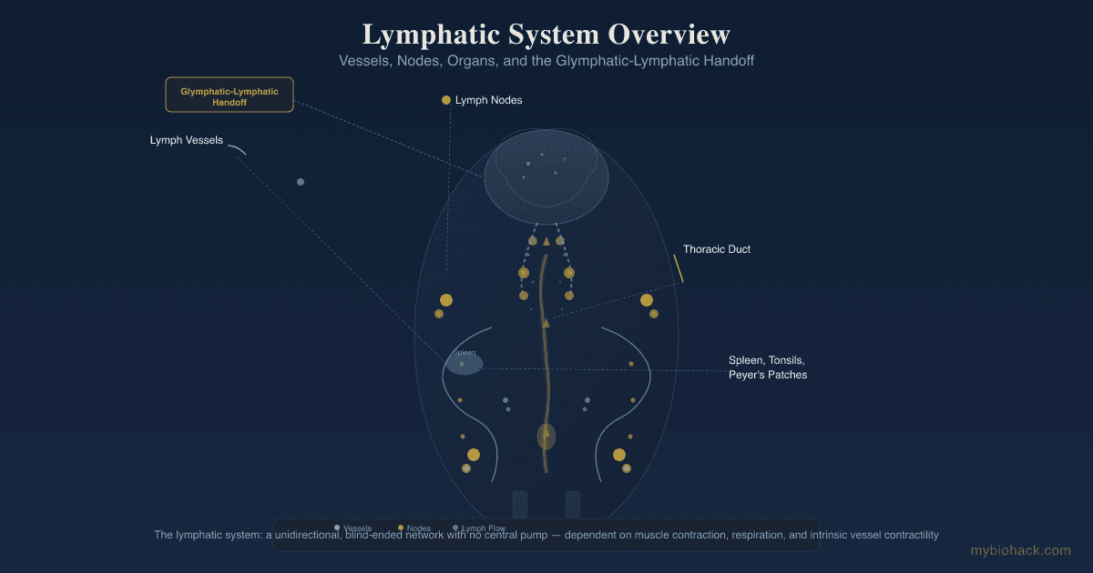
Lymphatic System Support: How To Improve Lymph Flow And Drainage
By Jacob Gordon, INHC, FMT-CThis article contains affiliate links. As an Amazon Associate, MyBioHack earns from qualifying purchases at no extra cost to you. We only link products we research and stand behind.
The lymphatic system is a blind-ended, unidirectional network that depends on mechanical forces rather than a central pump, making it uniquely vulnerable to stagnation in chronic illness.
In this post, we will discuss the basics of lymphatic function, what causes lymph stasis, how lymphatic dysfunction overlaps with chronic conditions through the Junction Dysfunction framework, and practical protocols to restore lymph flow.
Basics Of The Lymphatic System
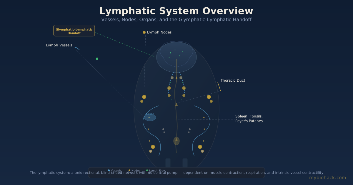
The lymphatic system is a network of vessels, nodes, and organs that runs parallel to the blood circulatory system.
Unlike the cardiovascular system, the lymphatic system has no central pump.
Lymph fluid moves through a combination of skeletal muscle contraction, respiratory pressure changes, arterial pulsation, and smooth muscle contraction within lymphatic vessel walls. R
The lymphatic system performs three primary functions: fluid balance by returning interstitial fluid to the bloodstream, immune surveillance by transporting antigens and immune cells through lymph nodes, and dietary fat absorption via intestinal lacteals.
Approximately 2 to 4 liters of lymph circulate through the body each day, and disruption of this flow is common in chronic illness.
The deep lymphatic network connects to the superficial lymphatic system via collecting vessels that pass through lymph nodes, where filtration and immune cell sampling occur.

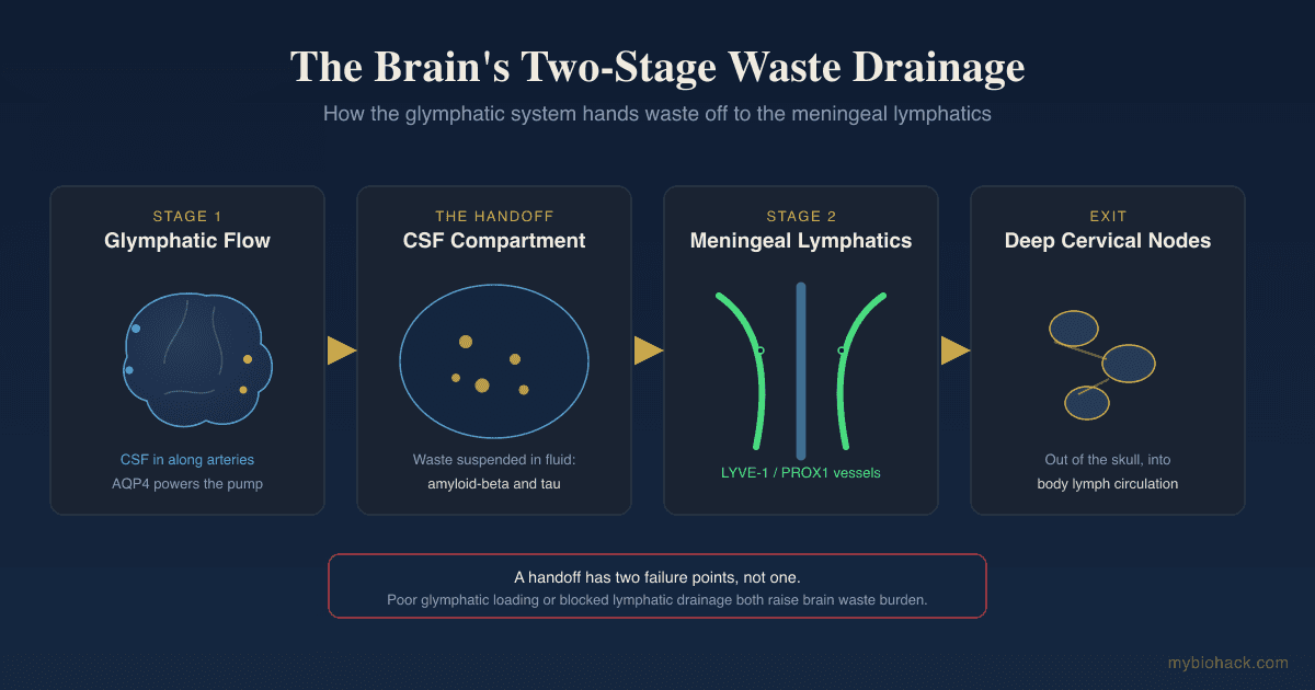
The glymphatic system is a parallel clearance network in the central nervous system that uses perivascular channels and Aquaporin-4 (AQP4) water channels on astrocytic endfeet to exchange cerebrospinal fluid with interstitial fluid, clearing metabolic waste like amyloid-beta and tau. R
The glymphatic and peripheral lymphatic systems are physically connected via meningeal lymphatic vessels that drain cerebrospinal fluid from the brain to deep cervical lymph nodes. R
This glymphatic-lymphatic handoff means that peripheral lymphatic dysfunction can impair brain waste clearance, and vice versa.
What Causes Lymph Stagnation
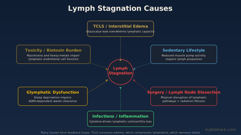
Lymph stagnation, also called lymphatic congestion or impaired lymph transport, occurs when the mechanical forces needed to move lymph are compromised or when the lymphatic vessels themselves are damaged. R
Physical Inactivity
Skeletal muscle contraction is the primary driver of lymph flow.
Sedentary behavior, bed rest, and immobilization reduce the muscle pump action that propels lymph through collecting vessels.
Inflammation And Fibrosis
Chronic inflammation damages lymphatic endothelial cells and collecting vessel valves.
Inflammatory cytokines like Tumor Necrosis Factor-Alpha (TNF-alpha) and Interleukin-1 beta disrupt lymphatic contractility and promote lymphangiogenesis that produces dysfunctional, leaky vessels. R
Fibrosis from repeated inflammation physically restricts lymphatic vessels, reducing their diameter and compliance.
Surgery And Lymph Node Dissection
Lymphadenectomy (lymph node removal) for cancer staging physically disrupts lymphatic pathways.
Radiation therapy causes lymphatic fibrosis that can persist for years after treatment ends. R
Obesity
Adipose tissue produces inflammatory mediators that impair lymphatic pump function.
Excess visceral fat physically compresses lymphatic collecting vessels, reducing flow.
Obesity is associated with reduced lymphatic contractility and increased interstitial fluid load. R
TCLS And Interstitial Edema
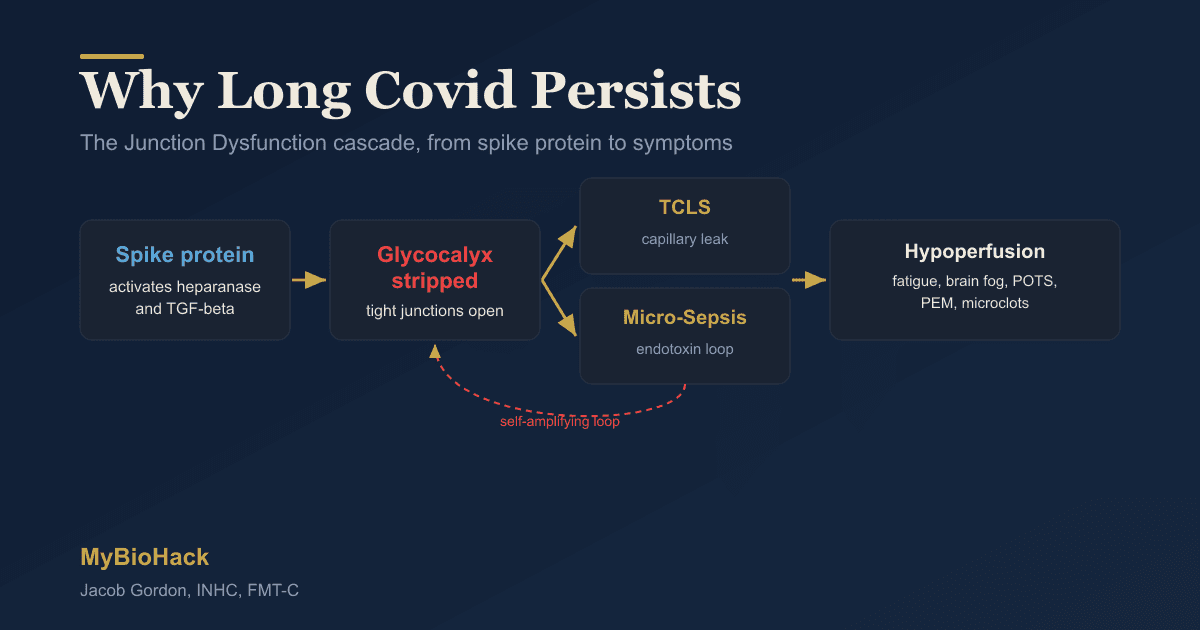
I coined Transient Capillary Leak Syndrome (TCLS) to describe the microvascular fluid leak that occurs when the glycocalyx becomes compromised.
TCLS forces fluid from the intravascular space into the interstitial space, overwhelming the lymphatic system's capacity to return it to circulation.
The resulting interstitial edema compresses initial lymphatic capillaries, reducing their ability to take up fluid.
This creates a feedback loop: TCLS causes interstitial fluid overload, which impairs lymphatic drainage, which worsens interstitial edema.
Glymphatic Dysfunction
Sleep deprivation, sympathetic dominance, and AQP4 mislocalization all impair glymphatic clearance.
Poor sleep is one of the strongest predictors of glymphatic dysfunction, since glymphatic flow increases significantly during sleep compared to wakefulness. R
When the glymphatic system cannot clear metabolic waste, the resulting neuroinflammation activates systemic inflammatory pathways that also impair peripheral lymphatic function. R
Dehydration
Lymph is approximately 95% water.
Chronic low water intake increases lymph viscosity, making it harder to move through narrow lymphatic capillaries.
Tight Clothing And Restricted Breathing
Compression of the thoracic and abdominal cavities from tight clothing, restrictive belts, or shallow breathing reduces the respiratory pump that pulls lymph upward through the thoracic duct.
The thoracic duct relies on negative intrathoracic pressure during inspiration to draw lymph into the venous circulation.
Shallow breathing from chronic stress, poor posture, or restrictive clothing reduces this pressure gradient.
Lymphatic And Overlapping Conditions
Post-Viral Syndromes (Long COVID, ME/CFS)
Post-viral illness disrupts the glymphatic-lymphatic axis through at least three mechanisms: glycocalyx degradation from spike protein exposure, autonomic dysfunction that reduces lymphatic pumping, and chronic inflammation that damages lymphatic vessels. R
Many symptoms attributed to "brain fog" in long COVID may represent glymphatic clearance failure.
I discuss this in more detail in the post on improving glymphatic function.
Mast Cell Activation Syndrome (MCAS)
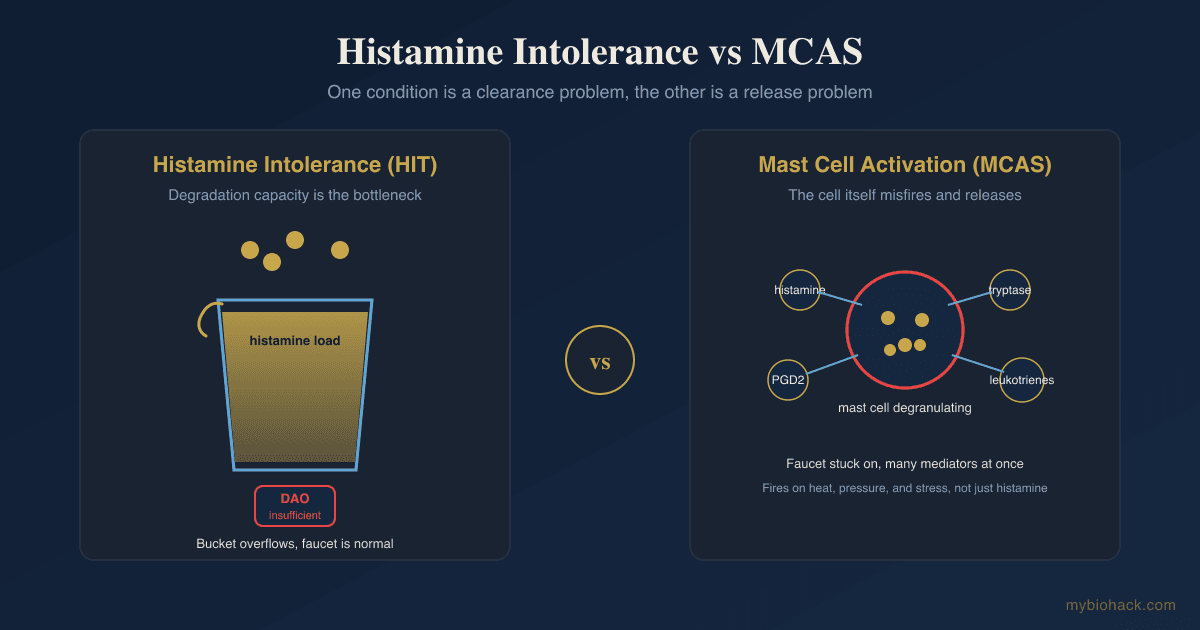
Mast cells cluster around lymphatic vessels and lymph nodes.
When mast cells degranulate, they release histamine, tryptase, and prostaglandins that alter lymphatic contractility and increase vascular permeability.
This is bidirectional: lymphatic stasis itself promotes mast cell degranulation in surrounding tissues.
For more detail, see the post on mast cells and substance P.
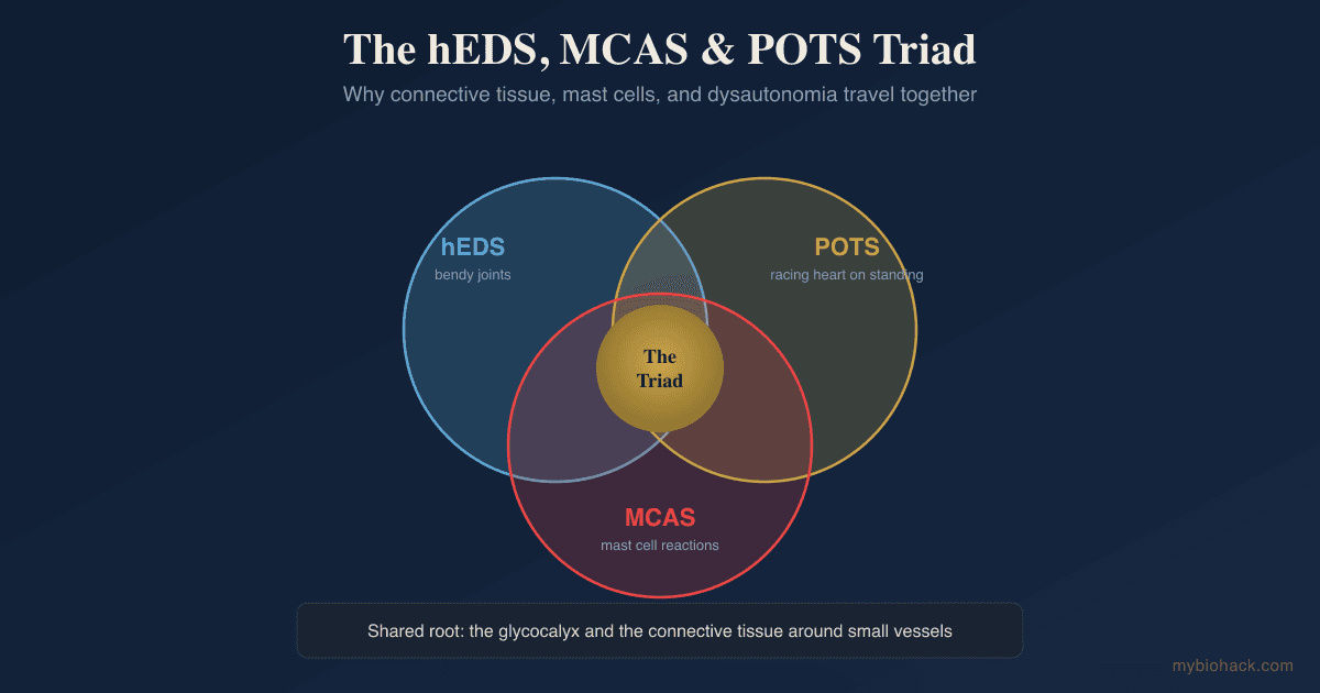
Ehlers-Danlos Syndrome / Hypermobility Spectrum Disorders
Connective tissue disorders affect the structural integrity of lymphatic vessels.
Collagen type 3 deficiencies in vascular EDS directly compromise lymphatic vessel wall strength.
Many hypermobile patients have impaired lymphatic flow that contributes to edema, brain fog, and immune dysfunction.
Neurodegenerative Diseases (Alzheimer's, Parkinson's)
Glymphatic clearance failure is a core mechanism in tau and alpha-synuclein accumulation. R
Peripheral lymphatic dysfunction reduces the clearance capacity of meningeal lymphatics, further impairing brain waste removal.
This is covered in the post on meningeal lymphatics.
Chronic Venous Insufficiency
Venous hypertension increases capillary filtration, adding to the interstitial fluid load that the lymphatic system must handle.
When lymphatic transport capacity is exceeded, venous edema transitions to phlebolymphedema. R
POTS / Dysautonomia
Autonomic dysfunction disrupts the sympathetic innervation of lymphatic smooth muscle.
Lymphatic vessels contract in response to both intrinsic (pacemaker) and extrinsic (neural) signals.
Sympathetic dominance impairs the parasympathetic-mediated relaxation phase that allows lymphangions to fill.
CIRS / Biotoxin Illness
Mycotoxins and other biotoxins directly impair lymphatic endothelial cell function.
Lymphatic congestion is common in mold illness and contributes to the inflammatory burden.
How To Improve Lymph Flow
1. Movement And Exercise
Muscle contraction is the most powerful driver of lymph flow.
Rhythmic, low-impact movement produces sustained lymph propulsion without the inflammatory burden of high-intensity exercise that can worsen glycocalyx damage in the JD population.
Walking: Brisk walking activates the calf muscle pump, which drives venous and lymphatic return from the lower extremities.
Walking for 20 to 30 minutes daily significantly improves lower extremity lymphatic clearance.
Rebounding: Mini-trampoline exercise (rebounding) creates gravitational acceleration changes that open and close lymphatic valves.
The up-and-down motion generates 2 to 3 times the gravitational force of walking, which mechanically stimulates lymph flow.
Deep breathing: Diaphragmatic breathing creates negative intrathoracic pressure that pulls lymph through the thoracic duct into the subclavian vein.
Ten slow, deep belly breaths performed several times per day can significantly enhance lymphatic return.
2. Manual Lymphatic Drainage (MLD)
MLD is a specialized light-pressure massage technique that follows the direction of lymphatic flow.
Unlike deep tissue massage, MLD uses gentle, rhythmic strokes (approximately 20 to 30 mmHg pressure) designed to stretch the skin and stimulate the lymphatic capillaries just beneath it.
A 2024 review noted that MLD evidence is strongest for mild lymphedema and early-stage intervention, with mixed results for advanced-stage disease. R
MLD has been shown to reduce edema and improve quality of life in patients with mild lymphedema, but the evidence is less conclusive for moderate to severe cases. R
For DIY approaches, self-MLD techniques are widely taught by certified lymphedema therapists.
Focus on the neck, axillary, and inguinal lymph node basins first to clear the "drainage pipes" before moving to the congested area.
3. Dry Brushing
Dry brushing uses a soft-bristle brush on dry skin in long, upward strokes toward the heart.
The evidence for dry brushing is largely anecdotal and mechanistic rather than clinical trial-based.
Dry brushing likely stimulates lymph flow through gentle mechanical stretching of initial lymphatic capillaries, similar to MLD but less precise.
The primary benefit may be exfoliation combined with increased cutaneous blood flow rather than direct lymphatic pumping.
If you try it, brush toward the heart, avoid broken skin, and do not brush aggressively.
4. Compression Therapy
Graduated compression stockings apply external pressure that reduces capillary filtration and supports lymphatic return.
Compression is the standard of care for lymphedema management in conventional medicine. R
For subclinical lymphatic congestion (puffiness, mild edema), 15 to 20 mmHg over-the-counter compression stockings may provide meaningful support.
Higher grades (20 to 30 mmHg, 30 to 40 mmHg) require a prescription and fitting by a certified fitter.
Alternating compression devices (pneumatic pumps) are also used for more advanced lymphatic insufficiency.
5. Hydration
Adequate water intake reduces lymph viscosity and improves flow through narrow lymphatic capillaries.
Electrolyte balance matters because sodium gradients influence interstitial fluid movement.
There is no single water target that fits everyone, but urine color in the pale yellow range is a reasonable daily hydration marker.
6. Sauna And Heat Therapy
Passive heating increases heart rate, cardiac output, and peripheral blood flow, all of which secondarily drive lymph flow.
Finnish sauna use (175 to 200 degrees Fahrenheit for 15 to 20 minutes, 4 to 7 times per week) is associated with reduced cardiovascular mortality in large Finnish cohort studies. R
Both traditional and infrared sauna increase sweating, which is itself a form of fluid and toxin elimination that reduces the burden on the lymphatic system.
7. Vibration Therapy
Whole-body vibration platforms mechanically stimulate lymphatic vessels through rapid oscillation.
The mechanical oscillation at 15 to 30 Hz stretches lymphatic initial capillaries and stimulates lymphangion contraction.
Small studies show vibration therapy reduces lower extremity edema in venous and lymphatic insufficiency.
8. Herbal Support
Red Root (Ceanothus americanus): A traditional lymphagogue used in Native American and eclectic medicine for swollen lymph nodes and splenic congestion.
Red root contains ceanothine alkaloids and tannins that are thought to stimulate lymphatic contractility and reduce tissue congestion.
Cleavers (Galium aparine): A classic alterative herb used for lymphatic swelling, skin conditions, and urinary support.
A 2020 study confirmed cleavers has immunostimulatory and antioxidant activity in vitro, partially validating its traditional use for lymphatic inflammation. R
Cleavers contains flavonoids (quercetin derivatives), iridoid glycosides, and tannins that support immune function and fluid elimination.
Parsley (Petroselinum crispum): A diuretic herb that supports renal elimination of metabolic waste, reducing the workload on the lymphatic system.
Parsley is rich in apigenin and myristicin, compounds with anti-inflammatory and diuretic properties.
9. Proteolytic Enzymes
Bromelain: A proteolytic enzyme complex derived from pineapple stem that reduces edema, inflammation, and fibrin deposition.
Bromelain breaks down fibrin and other proteins that accumulate in congested lymphatic tissue, improving fluid drainage.
A 2024 review confirmed bromelain's anti-edematous effects through modulation of NF-kB signaling and reduced pro-inflammatory cytokine production. R
Nattokinase: A fibrinolytic enzyme that degrades fibrin clots and improves blood and lymph viscosity.
Nattokinase may reduce the proteinaceous debris that accumulates in stagnant lymph.
10. Short-Chain Fatty Acids
Butyrate and other Short-Chain Fatty Acids (SCFAs) produced by gut microbiota fermentation of resistant starch support lymphatic integrity.
SCFAs regulate the differentiation of lymphatic endothelial cells and maintain intestinal lacteal integrity.
I cover this in detail in the post on short-chain fatty acids.
11. Quercetin
Quercetin stabilizes mast cells and reduces histamine release, which in turn reduces vascular permeability and interstitial fluid load.
Quercetin also inhibits MMP-9, an enzyme that degrades the glycocalyx and lymphatic basement membranes. R
For a full discussion, see the post on quercetin for mast cell and vascular health.
12. Sleep Optimization
Glymphatic clearance is significantly higher during sleep than wakefulness. R
Sleep position matters: lateral (side) sleeping improves glymphatic clearance compared to supine or prone.
Consistent sleep timing reinforces the circadian rhythm of AQP4 polarization and glymphatic flow. R
What To Stay Away From
Prolonged sitting: Staying seated for more extended periods significantly impairs lower extremity lymph return.
Stand up and walk for a few minutes every hour to reactivate the calf muscle pump.
Tight, restrictive clothing: Bras with underwire, tight waistbands, and compression at the groin or axilla physically obstruct lymphatic pathways.
Switch to wire-free or well-fitted bras, and avoid clothing that leaves red marks or indentations.
Dehydrating beverages: Caffeine and alcohol are diuretics that can dehydrate tissues and increase lymph viscosity if consumed in excess.
Moderate intake (1 to 2 cups of coffee or 1 alcoholic drink) is generally fine for most people, but chronic high intake impairs lymphatic flow.
High sodium processed foods: Excessive sodium increases interstitial fluid volume through osmotic retention.
Processed foods with hidden sodium contribute to the fluid load the lymphatic system must manage.
Deep tissue massage on congested areas: Aggressive massage on swollen or congested lymphatic tissue can damage initial lymphatic capillaries and worsen inflammation.
Always clear proximal lymph nodes first and use light pressure when working on lymphatic areas.
Deconditioning / overtraining: Both extremes impair lymphatic function.
Sedentary behavior reduces muscle pump activity, while excessive high-intensity exercise increases inflammation and glycocalyx shedding without allowing adequate recovery.
Chronic sleep deprivation: Glymphatic clearance depends on adequate sleep.
Consistent sleep loss creates a backlog of metabolic waste that the glymphatic system cannot clear. R
Testing
Imaging
Lymphoscintigraphy: The gold standard imaging modality for evaluating lymphatic function.
A radioactive tracer is injected into the interstitial space, and its movement through lymphatic vessels is tracked over time.
Delayed or absent tracer transit indicates lymphatic obstruction or pump failure.
Near-Infrared Fluorescence Lymphatic Imaging: Uses indocyanine green (ICG) dye injected intradermally, followed by near-infrared camera imaging.
ICG lymphography provides real-time visualization of superficial lymphatic architecture and is increasingly used in clinical lymphatic assessment.
Duplex Ultrasound: Useful for evaluating venous insufficiency, which frequently coexists with lymphatic dysfunction.
Venous reflux or obstruction increases the interstitial fluid load that lymphatics must manage.
Blood And Urine Markers
TNF-alpha: a pro-inflammatory cytokine that impairs lymphatic contractility when chronically elevated.
High-Sensitivity C-Reactive Protein (hsCRP): systemic inflammation marker that correlates with lymphatic dysfunction.
MMP-9: matrix metalloproteinase that degrades the glycocalyx and lymphatic basement membranes.
Fasting insulin and HOMA-IR: insulin resistance promotes lymphatic endothelial dysfunction and reduces lymphatic pumping capacity.
Functional Lab Panels
I use the Immune Zoomer (Vibrant Wellness) to assess mast cell mediators, autoantibodies, and immune reactivity that may drive lymphatic inflammation.
I use the Cardio Zoomer (Vibrant Wellness) to evaluate endothelial function markers, including MMP-9, vascular inflammation, and metabolic contributors to lymphatic dysfunction.
I use the Toxin Zoomer (Vibrant Wellness) to assess mycotoxin and heavy metal burden, both of which impair lymphatic endothelial function.
I use the Cellular Zoomer (Vibrant Wellness) to evaluate oxidative stress and mitochondrial function, which influence lymphatic smooth muscle contraction.
I use the Gut Zoomer (Vibrant Wellness) to assess intestinal permeability and dysbiosis, both of which contribute to systemic inflammation that impairs lymphatic function.
Provocation Testing
Exercise challenge: Evaluate for increased swelling or heaviness in the limbs after 15 to 20 minutes of walking or standing.
Worsening symptoms with activity suggest mechanical or obstructive lymphatic insufficiency rather than pump failure.
Elevation test: If limb swelling reduces significantly after 24 hours of leg elevation (above heart level), the lymphatic system still has residual transport capacity.
If swelling does not reduce with elevation, the lymphatic system has more advanced structural impairment.
Mechanisms Of Action
Simple:
Lymphatic vessels contract rhythmically like a series of tiny hearts, pushing fluid forward through one-way valves.
When you contract your muscles, they squeeze these vessels and propel lymph toward the heart.
Deep breathing creates a vacuum that pulls lymph upward through the chest.
Any intervention that increases rhythmic muscle contraction, respiratory motion, or gentle mechanical stretching of the skin will improve lymph flow.
Advanced:
- Lymphangion Pacemaker Activity: Each lymphangion (the segment of a lymphatic vessel between two valves) contains smooth muscle that contracts autonomously in response to calcium oscillations in lymphatic endothelial cells. R
Contraction frequency is modulated by nitric oxide (NO) production, with low NO promoting contraction and high NO promoting relaxation.
Inflammatory cytokines like TNF-alpha disrupt this NO balance, impairing the contraction-relaxation cycle needed for forward propulsion.
- Calf Muscle Pump And Venous-Lymphatic Coupling: Contraction of the gastrocnemius and soleus muscles compresses both deep veins and lymphatic collecting vessels, generating pressures of 150 to 200 mmHg.
This compression propels blood toward the heart and lymph toward the subclavian vein simultaneously.
The lymphatic and venous systems share anatomical pathways and respond synergistically to muscle contraction. R
- Respiratory Pump: During inspiration, the diaphragm descends, increasing intra-abdominal pressure and decreasing intrathoracic pressure.
This pressure gradient creates a suction effect that draws lymph from the cisterna chyli through the thoracic duct into the left subclavian vein.
Diaphragmatic excursion during deep breathing generates a pressure swing of 5 to 15 mmHg, significantly enhancing thoracic duct flow.
- Glycocalyx-Lymphatic Integrity: The endothelial glycocalyx on lymphatic collecting vessels regulates permeability and mechanotransduction.
Glycocalyx degradation (as in JD) impairs the ability of lymphatic endothelium to sense shear stress and respond with appropriate contraction.
AQP4 polarization on astrocytic endfeet in the glymphatic system depends on the perivascular glycocalyx, and when the glycocalyx is lost, AQP4 becomes delocalized and glymphatic flow drops. R
This is detailed in the post on the interstitium, which covers the fluid dynamics of the body's third circulatory system.
- S1P Signaling And Lymphatic Endothelial Barrier: Sphingosine-1-phosphate (S1P) signaling through S1PR1 on lymphatic endothelial cells maintains junctional integrity and regulates lymphocyte trafficking. R
Loss of S1P signaling in lymphatic endothelial cells worsens lymphatic insufficiency and promotes pathogenic CD4 T cell infiltration.
This pathway is a target for emerging lymphatic therapeutics.
- FOXC2 And Lymphatic Valve Maintenance: The forkhead transcription factor FOXC2 controls the formation and maintenance of lymphatic valves through cooperation with NFATc1. R
Oscillatory shear stress from lymph flow upregulates FOXC2, which in turn regulates connexin 37, ephrinB2, and other valve-related genes. R
Loss of FOXC2 function leads to valve agenesis, lymph reflux, and progressive lymphatic insufficiency. R
- Mechanotransduction And PROX1 Regulation: Lymphatic endothelial cells sense fluid flow through primary cilia and glycocalyx-mediated mechanosensors.
Flow-induced shear stress upregulates PROX1 (the master regulator of lymphatic identity) and FOXC2 through calcium-calcineurin-NFAT signaling.
This mechanosensory feedback loop maintains lymphatic vessel identity and function as long as flow is present.
When flow stagnates, PROX1 and FOXC2 expression falls, initiating a positive feedback loop of progressive dedifferentiation and further stagnation.
Genetics
FOXC2
The FOXC2 gene encodes a forkhead transcription factor essential for the development and maintenance of lymphatic and venous valves.
Mutations in FOXC2 cause lymphedema-distichiasis syndrome, a hereditary form of primary lymphedema characterized by lower extremity swelling and an extra row of eyelashes. R
More than 50 FOXC2 mutations have been identified, most of which produce a truncated, nonfunctional protein. R
FOXC2 haploinsufficiency is the most common cause of inherited lymphatic valve dysfunction.
VEGFC / VEGFR3
Vascular Endothelial Growth Factor C (VEGFC) and its receptor VEGFR3 are the primary signaling axis for lymphatic development.
Loss-of-function mutations in VEGFR3 (FLT4) cause hereditary lymphedema type 1 (Milroy disease), presenting at birth with lower extremity swelling.
VEGFC polymorphisms influence lymphangiogenic capacity and recovery from lymphatic injury.
GATA2
GATA2 is a transcription factor required for lymphatic valve development and maintenance.
GATA2 mutations cause MonoMAC syndrome, which includes primary lymphedema as a feature.
GATA2 cooperates with FOXC2 to regulate valve-specific genes during development. R
PROX1
PROX1 is the master regulator of lymphatic endothelial cell identity.
Prox1 expression distinguishes lymphatic endothelial cells from blood vascular endothelial cells during development.
Reduced PROX1 expression in adults leads to lymphatic dedifferentiation and loss of vessel identity, contributing to progressive lymphatic failure.
HGF / MET
Hepatocyte Growth Factor (HGF) signaling through the MET receptor promotes lymphangiogenesis and lymphatic vessel repair.
HGF/MET signaling is upregulated in response to lymphatic injury and promotes the sprouting of new lymphatic capillaries.
Polymorphisms in HGF and MET influence lymphatic repair capacity after surgery or radiation.
More Research
Glymphatic clearance is state-dependent and circadian.
Glymphatic flow peaks during the mid-rest phase (in humans, during early sleep), and this rhythm is lost in conditions of chronic sleep disruption. R
AQP4 polarization varies across the circadian cycle, with highest polarization during the rest phase and lowest during the active phase.
This means that sleep timing is at least as important as sleep duration for brain waste clearance.
CD4 T cell-driven lymphatic damage is a newly recognized mechanism.
Pathogenic Th1, Th2, and Th17 cells infiltrate lymphatic vessels after injury and release cytokines that impair contractility and promote fibrosis. R
P-selectin inhibition and S1PR1 restoration are emerging therapeutic targets currently in preclinical development.
Lymphatic system dysfunction contributes to heart failure progression.
In chronic heart failure, cardiac lymphatic remodeling is insufficient to handle the increased interstitial fluid load, and this worsened lymphatic dysfunction contributes to edema and inflammation. R
Restoring cardiac lymphatic drainage is being investigated as a therapeutic target. R
The Long COVID Natural Treatment Protocol covers how lymphatic and microvascular dysfunction interact in post-viral recovery.
For a deeper mechanistic understanding, the Junction Dysfunction guide covers Chapter 35 (Lymph / Glymphatic) with the full cascade of glycocalyx-driven interstitial stasis and lymphatic pump failure.
For biomarker testing I use the Immune Zoomer for inflammatory profiles and the Cardio Zoomer for endothelial and vascular markers relevant to lymphatic dysfunction.
Jacob Gordon
INHC, FMT-C
Integrative Nutrition Health Coach
I cover mold illness, post-viral recovery, methylation, and complex chronic disease, drawing on ten years of clinical research, work inside a functional medicine clinic, and my own recovery from all of it. Every claim here is cited.
Book a ConsultationRelated Protocols & Supplements
Deep-dive chapters and recommended supplements for this topic
Electrolyte Complex
1 scoop/day
CoQ10
200mg/day
Magnesium Glycinate
400mg at bedtime